Figure 4. Representative photomicrographs of double immunostaining of the P2X7 receptor and CD68 in the normal retina and in the retina on days 1, 2, 3, and 7 after intraocular pressure elevation. P2X7-positive cells were also stained with anti-CD68 in the ganglion cell layer (GCL). Bar=100 µm.
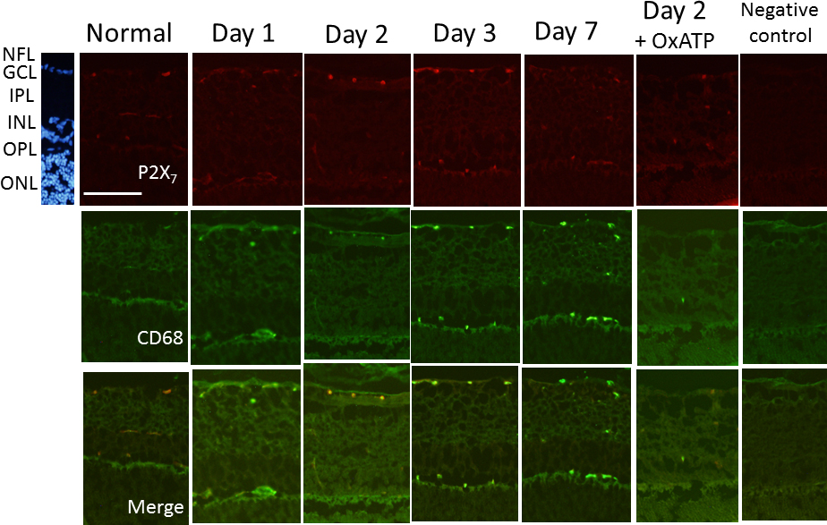
 Figure 4 of
Sugiyama, Mol Vis 2013; 19:2080-2091.
Figure 4 of
Sugiyama, Mol Vis 2013; 19:2080-2091.