Figure 2. Effects of the P2X7 antagonists—oxidized adenosine triphosphate (3–100 µM) and brilliant blue G (0.3–300 nM)—on the retinal changes after IOP
elevation. A: Cell density in the ganglion cell layer (GCL), B: inner plexiform layer (IPL) thickness. Data are expressed as the mean±standard error of the mean (SEM; n=4–8). The cell
density in the eyes injected with phosphate-buffered saline (PBS) after intraocular (IOP) elevation was significantly lower
than that in the sham control eyes (unpaired Student t test, p<0.01 for A, p<0.05 for B). The asterisks indicate significant differences in eyes after IOP elevation and intravitreal injection of PBS (unpaired
Student t test, **p<0.01, *p<0.05).
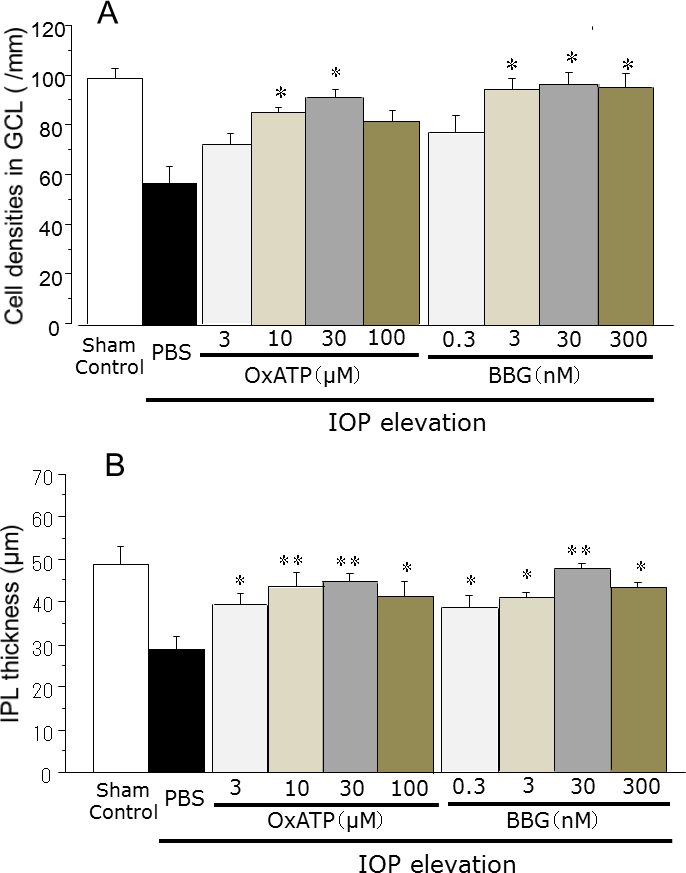
 Figure 2 of
Sugiyama, Mol Vis 2013; 19:2080-2091.
Figure 2 of
Sugiyama, Mol Vis 2013; 19:2080-2091.