Figure 1. Photomicrographs of transverse sections of the posterior retina stained with hematoxylin and eosin. Sections were obtained
from a sham control eye (A) and from eyes after intraocular pressure (IOP) elevation and intravitreal injection of phosphate-buffered saline (PBS; B) or the P2X7 antagonists—oxidized adenosine triphosphate (OxATP; 30 µM; C) and brilliant blue G (BBG; 30 nM; D). Cell loss in the ganglion cell layer (GCL) and thinning of the inner plexiform layer (IPL) were observed in the eyes after
IOP elevation and PBS, but were rescued by P2X7 antagonists. Bar=100 µm.
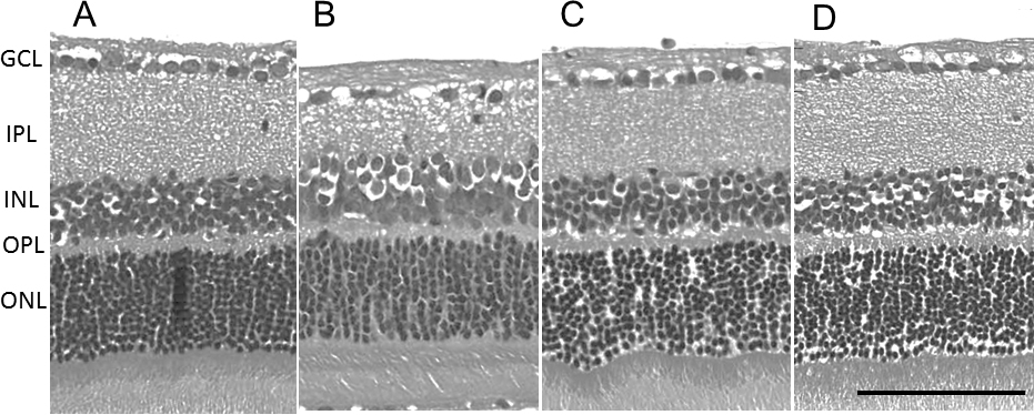
 Figure 1 of
Sugiyama, Mol Vis 2013; 19:2080-2091.
Figure 1 of
Sugiyama, Mol Vis 2013; 19:2080-2091.