Figure 3. Pcdh21 staining occurs in photoreceptors in the retina of postnatal day 4 and adult mice expressing green fluorescent protein
under the control of the neural retina leucine zipper promoter. Green fluorescent protein is expressed in developing (B) and mature (F) rod photoreceptors. Pcdh21 is localized at the tip of the developing inner segment in the retina (A, C, D) of 4-day-old mice. In the adult retina, Pcdh21 staining was observed at the connecting cilium between the inner and outer
segments (E, G, H). D and H show magnifications of C and G. Scale bar in C representative for A and B and scale bar in G representative for E and F: 50 μm. Scale bars in D and H: 10 μm. INL: inner nuclear layer; IS: inner segments; NBL: neuroblast layer; ONL: outer nuclear layer; OS: outer segments;
RGC: retinal ganglion cell layer.
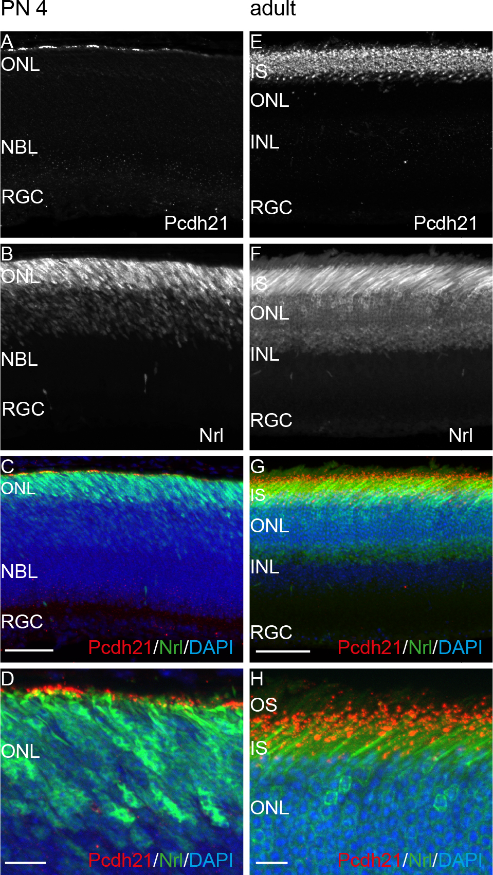
 Figure 3 of
Postel, Mol Vis 2013; 19:2058-2067.
Figure 3 of
Postel, Mol Vis 2013; 19:2058-2067.