Figure 7. Second harmonic generation images. Second harmonic generation (SHG) imaging shows collagen orientation in the sclera of an
older CD1 mouse; the 1 mm2 sclera piece includes the optic nerve canal (seen as the dark central area). The superior is located at the top of the images,
while blood vessels (*) surround the optic nerve head on the nasal, inferior and temporal portion of the sclera. A: The peripapillary (posterior) sclera in control specimen had collagen fibrils oriented in a circumferential fashion around
the optic nerve. B: In eyes exposed to elevated intraocular pressure (IOP), there was a retraction of this circumferential zone away from the
optic canal, especially in the superior area. The white boxes in first two images (A, B) highlight the superior region. C: A 20× image of the posterior sclera shows no retraction in control tissue at the circumferential zone of the superior region.
D: The retraction of collagen fibrils can be seen at the circumferential zone of the superior quadrant near the optic canal
in 6 week glaucoma-treated eyes (20X). Moreover, 40× images of the mid-sclera show collagen assembled in a basket weave formation,
where E: untreated tissue and F: glaucoma-treated tissue, which shows a slight change in fibril assembly. Scale bars are equal to 100µm for images A and B, and 10 µm for images C, D, E, and F.
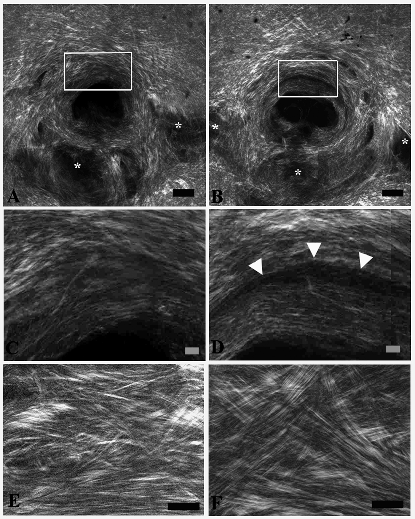
 Figure 7 of
Cone-Kimball, Mol Vis 2013; 19:2023-2039.
Figure 7 of
Cone-Kimball, Mol Vis 2013; 19:2023-2039.