Figure 4. Collagen fibril diameter distribution by sclera location in CD1 control. The distribution for 100 collagen fibrils from a
control CD1 mouse taken from the inner, mid-, and outer sclera shows that the mid-sclera has a broader range of fibril diameter
and higher mean fibril diameter. The inner and outer sclera were different when compared to the larger fibrils found in the
middle portion of the sclera (≤0.0001 and ≤0.0001 respectively, t test).The inner and outer scleral collagen diameter measurements, however, were not statistically different from one another.
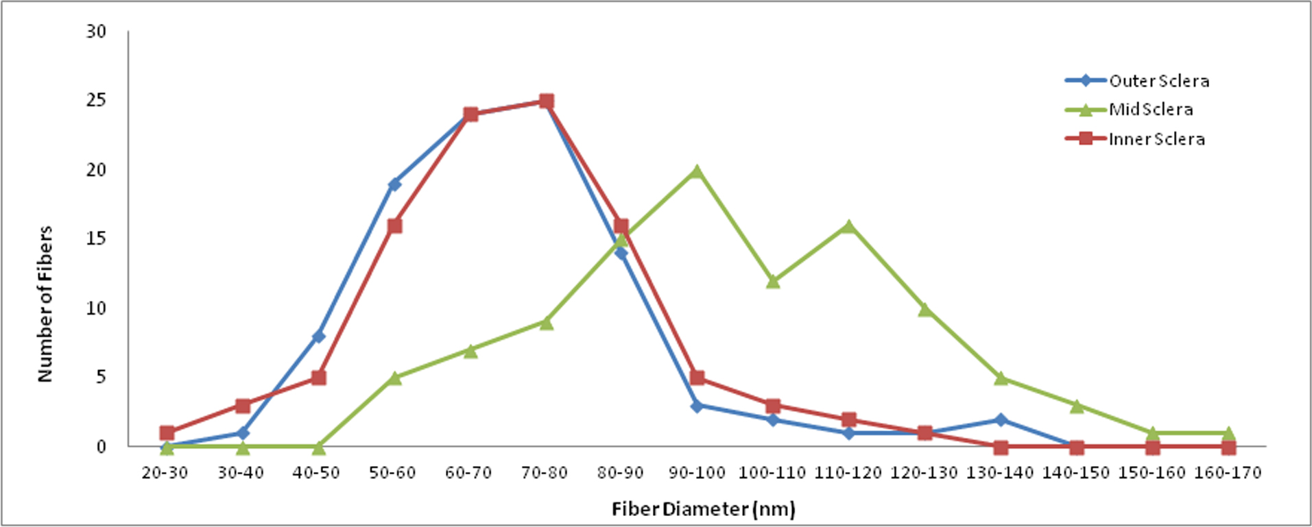
 Figure 4 of
Cone-Kimball, Mol Vis 2013; 19:2023-2039.
Figure 4 of
Cone-Kimball, Mol Vis 2013; 19:2023-2039.