Figure 3. Collagen lamella orientation
. Using transmission electron microscopy (TEM), the lamellae of the sclera were quantified and their thickness measured by
dividing them into the following four types based on their orientation of collagen fibrils: cross-section (fibrils anterior-posterior=CR),
longitudinal (parallel to equator, nasal-temporal=L), oblique (O), and lamellae consisting of sclera fibroblasts (cellular=CL;
scale bar=500 nm).
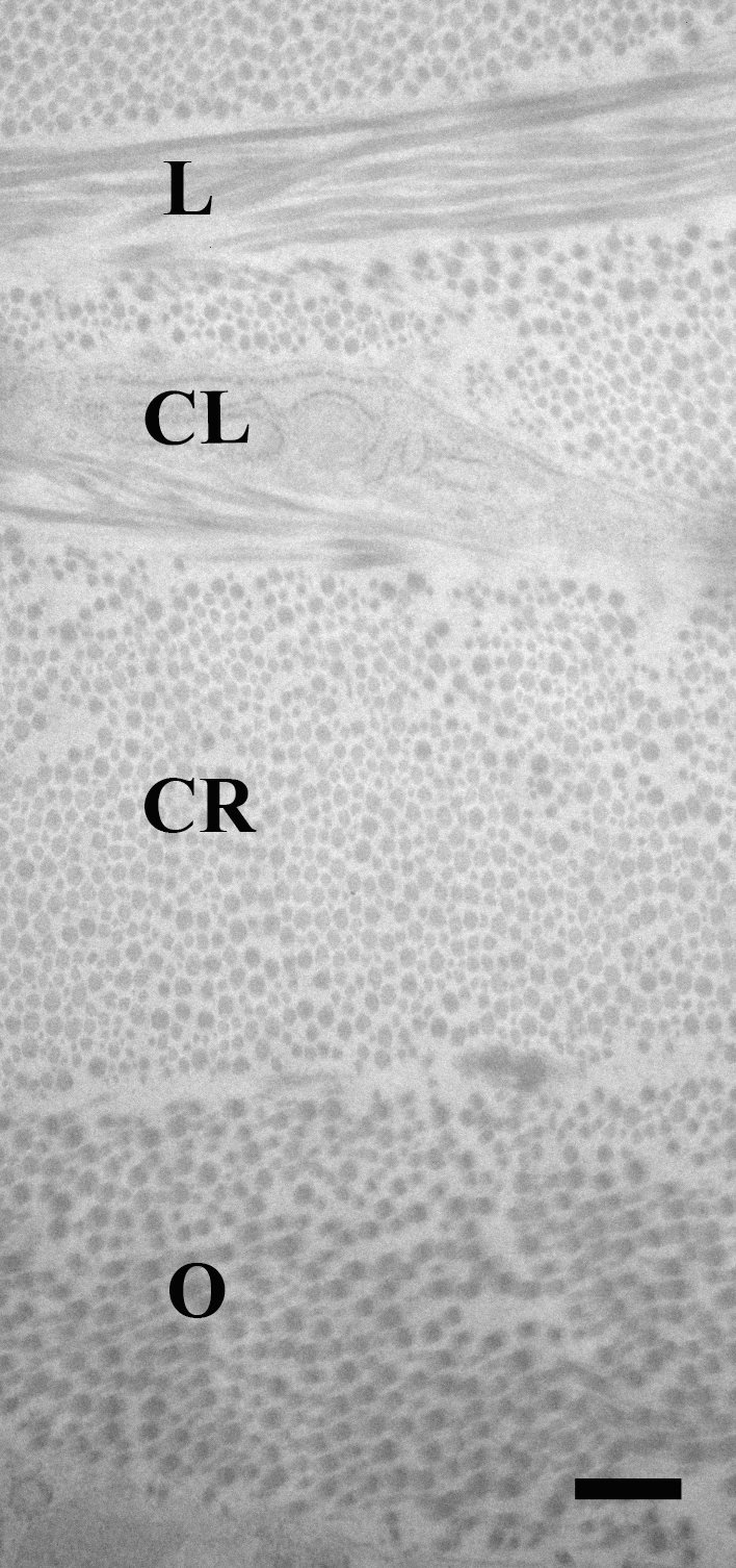
 Figure 3 of
Cone-Kimball, Mol Vis 2013; 19:2023-2039.
Figure 3 of
Cone-Kimball, Mol Vis 2013; 19:2023-2039.