Figure 2. Scleral tissue division. The method of identifying areas of the sclera for measurement of scleral thickness, lamellar orientation,
and fibril diameter distribution is shown. A: For sclera thickness measurements in fresh tissue slices, the sclera was divided into six regions, labeled R1 (peripapillary)
to R6 (limbus). B: The sclera was divided into the following pieces; (1) Four zones; superior, nasal, inferior, and temporal. (2) The optic
nerve head and peripapillary sclera were removed with a 1.5 mm in diameter hole punch. (3) Razor cuts were made at the superior
to assist with identification during plastic embedding and sectioning. (4–7) From each quadrant, two specimens (representing
Regions 2+3 and Region 4 in A above) were selected for epoxy embedding, as was the peripapillary specimen.
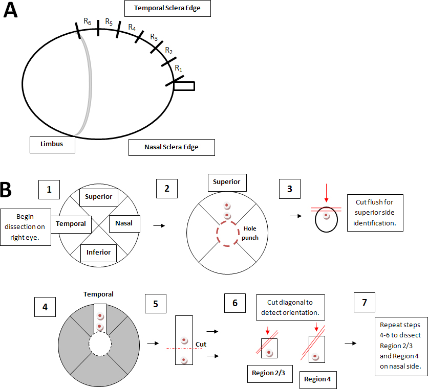
 Figure 2 of
Cone-Kimball, Mol Vis 2013; 19:2023-2039.
Figure 2 of
Cone-Kimball, Mol Vis 2013; 19:2023-2039.