Figure 4. Immunohistochemistry for inhibitor of apoptosis 1, the retinal ganglion cell marker Thy 1, glial fibrillary acidic protein,
and 4',6-diamidino-2-phenylindole in retinal cryosections of young and old rats at 8 days after induction of elevated intraocular
pressure (IOP). The merged image shows colocalization of IAP with Thy 1 (yellow) and with glial fibrillary acidic protein
(GFAP; purple), suggesting that the source for changes in IAP expression is from retinal ganglion cells (RGCs) and glial cells.
A: In 3-months-old rats, IAP levels increased in glaucomatous eyes as well as staining for GFAP. B: IAP-1 staining decreased in old glaucomatous 13-month-old eyes as compared to fellow eyes. Magnification 40X, scale bars:
all panels 20 μm.
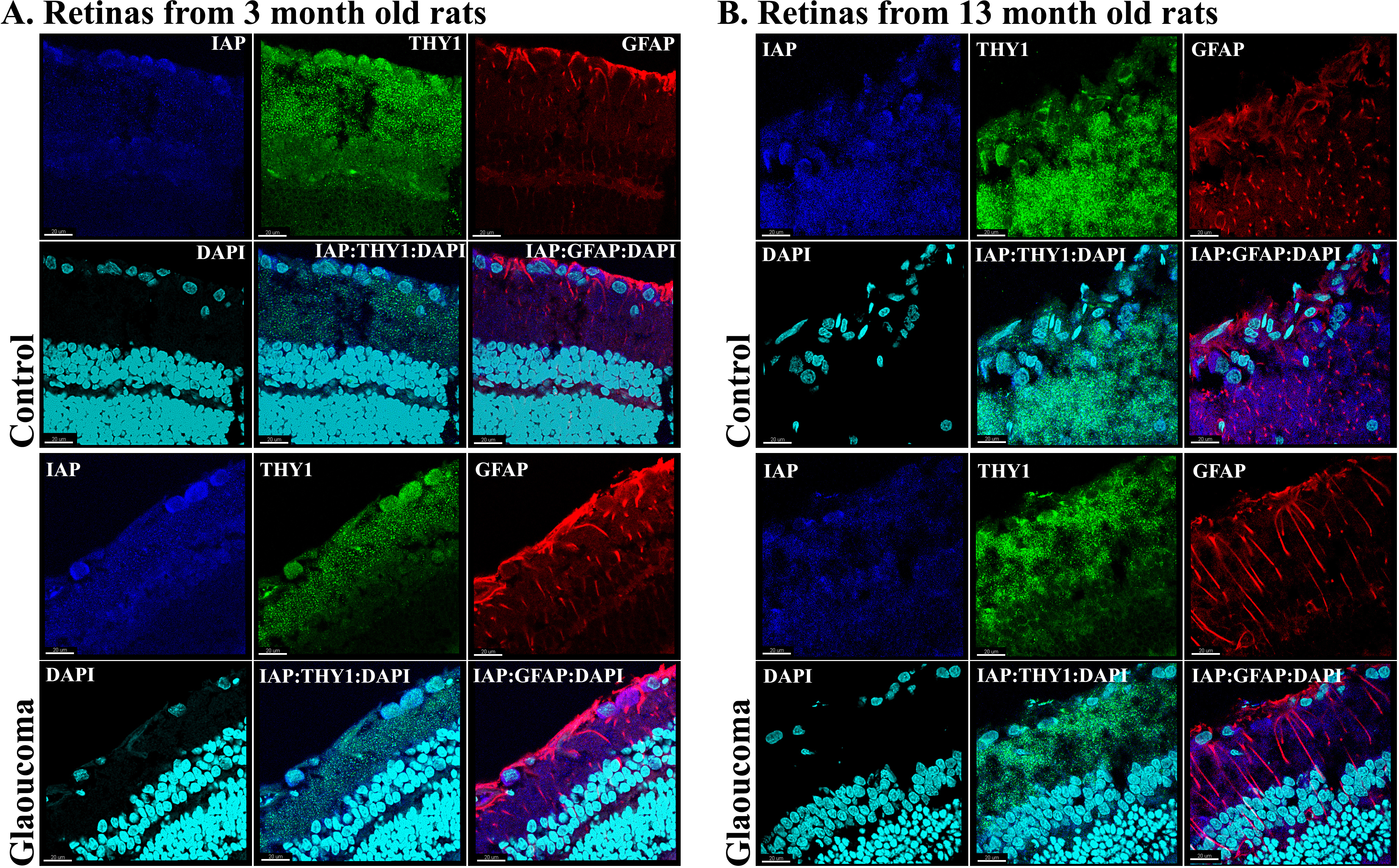
 Figure 4 of
Levkovitch-Verbin, Mol Vis 2013; 19:2011-2022.
Figure 4 of
Levkovitch-Verbin, Mol Vis 2013; 19:2011-2022.