Figure 2. Retinal ganglion cell loss increased with age in both glaucomatous and control fellow eyes. A: The mean retinal ganglion cell (RGC) survival 10 weeks after the induction of elevated intraocular pressure (IOP) is shown.
There was a significant decrease in RGCs in the control fellow eyes with age (n=4–8 for each age group, data presented as
SEM, p=0.002), as well as in the glaucomatous eyes (n=4–8, p=0.048). B: The amount of glaucomatous RGC loss increased with age (n=4–8, p=0.05). This progression in RGC loss due to age occurred
under similar IOP levels. C-H: Representative fluorogold images of RGCs 10 weeks after induction of glaucoma in young and old eyes are shown. Magnification
40X. I: Labeled RGCs were counted with a 40 super wide field objective along two radii in four directions (i.e., superior, temporal,
inferior, and nasal) centered on the position of the optic nerve head.
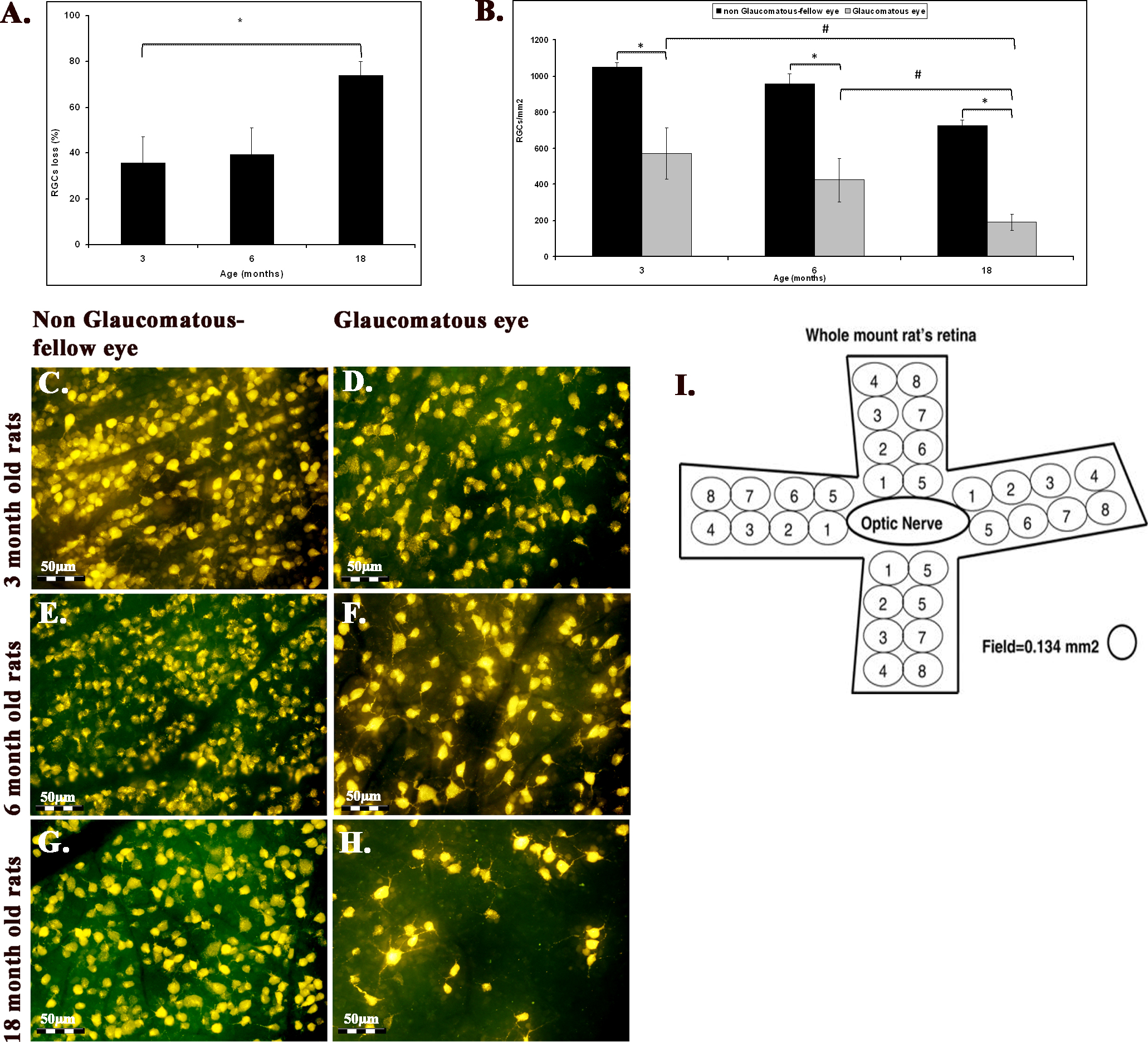
 Figure 2 of
Levkovitch-Verbin, Mol Vis 2013; 19:2011-2022.
Figure 2 of
Levkovitch-Verbin, Mol Vis 2013; 19:2011-2022.