Figure 3. The protective effect of total flavonoids from Flos Puerariae (TFF) on the ultrastructure of the retinal ganglion cells. n=4
per group. Five electrophotographs of each group were analyzed. A–E: The representative images show the ultrastructural changes in the retinal ganglion cells (RGCs) of each group (Scale bar=3
μm). F–J: The enlarged views of A–E (Scale bar=1 μm). A and F: In the control group, the RGCs had uniformly normal-appearing nuclei (N) and mitochondria (M). B and G: In the DM group, vacuolation (V), chromatin margination, chromatin condensation, swelling of mitochondria, and loss of cristae
were detected. C and H: In the 50 mg/kg TFF-treated group, there were still massive swollen mitochondria and chromatin condensation. D and I: In the 100 mg/kg TFF-treated group, mitochondria showed minor changes compared with the diabetic group, whereas chromatin
margination and condensation exist. E and J: In the 200 mg/kg TFF-treated group, mitochondria and nucleus were basic normal and occasional evidence of apoptosis was
detected. N: nucleus of the ganglion cell; M: mitochondria of the ganglion cell; V: vacuoles.
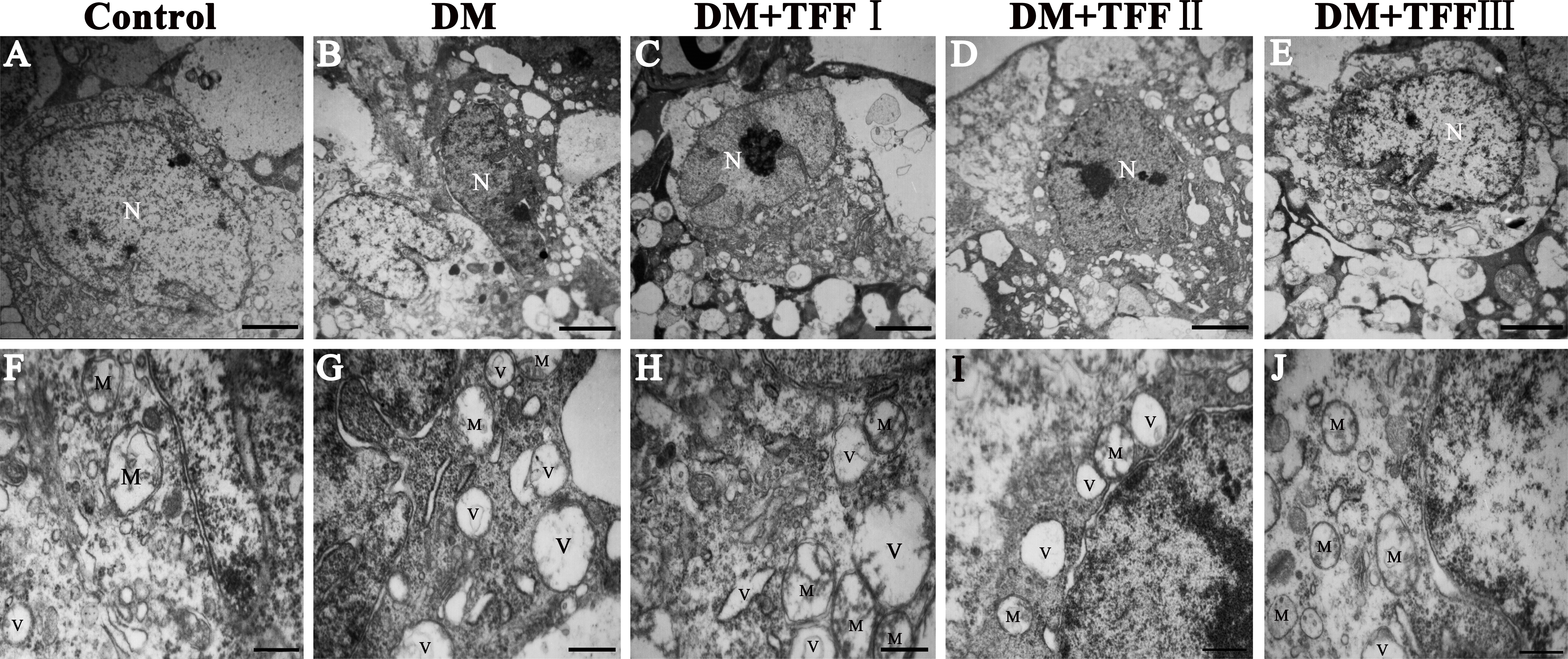
 Figure 3 of
Li, Mol Vis 2013; 19:1999-2010.
Figure 3 of
Li, Mol Vis 2013; 19:1999-2010.