Figure 1. Representative electrospray ionization tandem mass spectrometric analysis of sphingomyelins extracted from control human aqueous
humor in the positive-ion mode. A: Neutral loss scan (NLS) of m/z 213.2 corresponding to sphingomyelin class. B: NLS as above with internal standard addition (arrow head; m/z ratio of 729.1) enabling ratiometric quantification of all
identified lipids in each sphingomyelin class. The NLS for m/z 350–1000 is shown. C: Precursor ion scan (PIS) of m/z 184 corresponding to choline moiety within the sphingomyelins. D: PIS as above with internal standard addition (arrow head; m/z ratio of 729.1), enabling ratiometric quantification of all
identified lipids in sphingomyelin class using PIS. Thin arrows depict the indicated species. Appendix 2 is an enlarged version
of this figure.
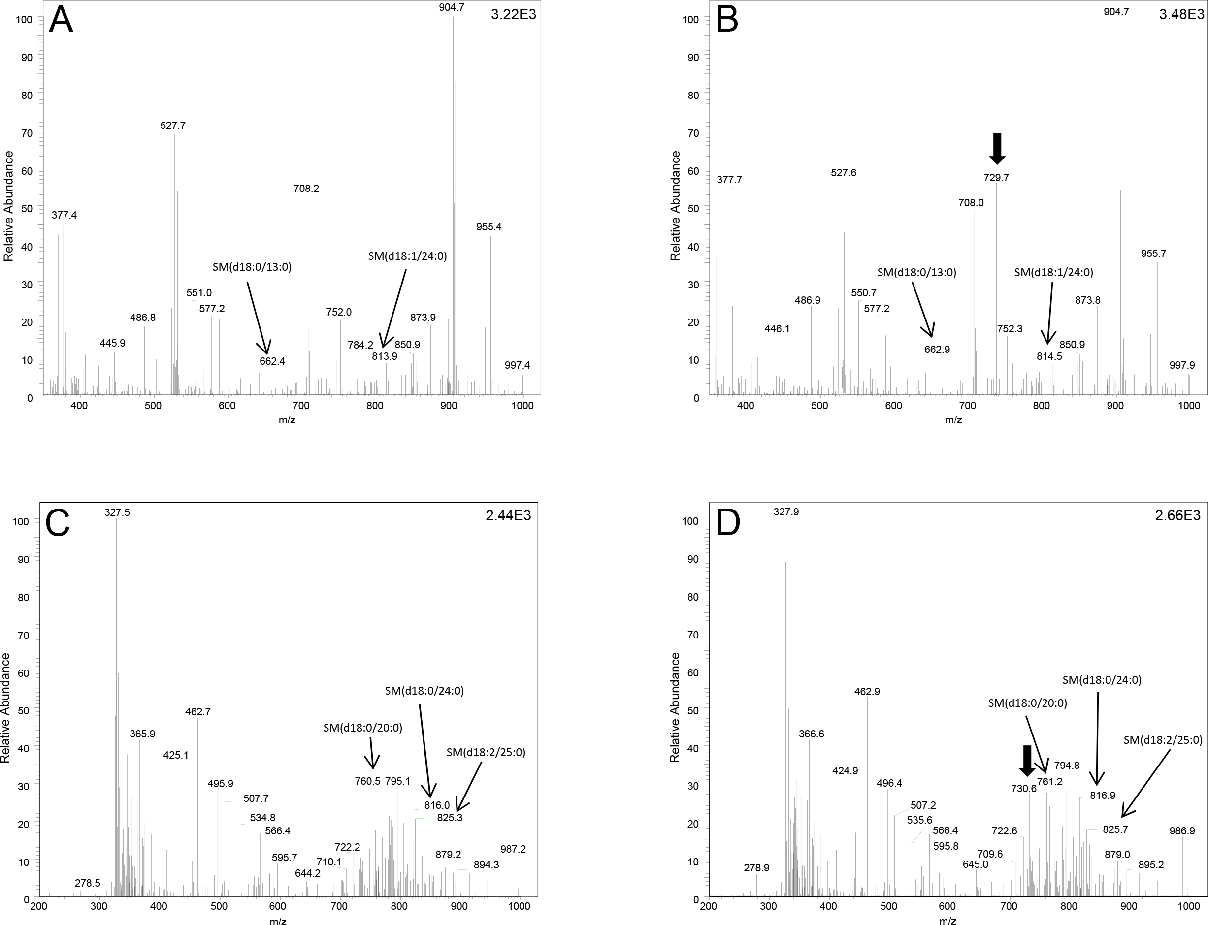
 Figure 1 of
Aljohani, Mol Vis 2013; 19:1966-1984.
Figure 1 of
Aljohani, Mol Vis 2013; 19:1966-1984.