Figure 1. Anakinra improved ocular surface integrity. A 14-day course of topical anakinra significantly decreased progression of lissamine
green staining in mice with autoimmune-mediated aqueous tear deficiency. A: Representative images of lissamine green staining for an autoimmune regulator (Aire) knockout (KO) mouse before (upper panels) and after (lower panels) 14-day treatment (Tx) with anakinra or carboxymethylcellulose
(CMC) vehicle control. Staining was significantly reduced in anakinra-treated eyes (lower right) compared to those treated
with vehicle (lower left). B: Lissamine green (LG) staining scores at days 7 and 14 are reported as a delta change relative to staining scores at baseline
(day 0). The change in LG staining following treatment at day 7 (day 7– day 0) was 0.5±0.90 (CMC vehicle) versus −0.5±0.35
(anakinra); p=0.28, where positive values represent increased LG staining and negative values indicate decreased staining.
By day 14 (day 14 – day 0), the change in LG reached 1.2±0.43 (CMC vehicle) versus −0.5±0.25 (anakinra); p=0.01).
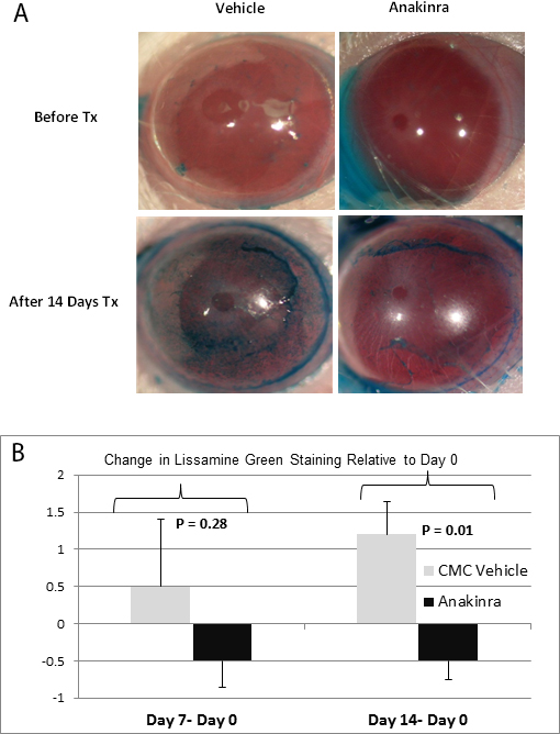
 Figure 1 of
Vijmasi, Mol Vis 2013; 19:1957-1965.
Figure 1 of
Vijmasi, Mol Vis 2013; 19:1957-1965.