Figure 1. Immunocytochemical images of cultured retinal ganglion cells. Positive retinal ganglion cells (RGCs) were stained green with
Thy-1.1, and their nuclei were stained blue with Hoechst 33342. A: Fluorescence microscope image of the retinal ganglion cell after labeling with Thy-1.1 (in green). B: Fluorescence microscope image of the retinal ganglion cell after labeling with Hoechst 33342 (in blue). C: Merged image of (A) and (B). Bar: 50 μm.
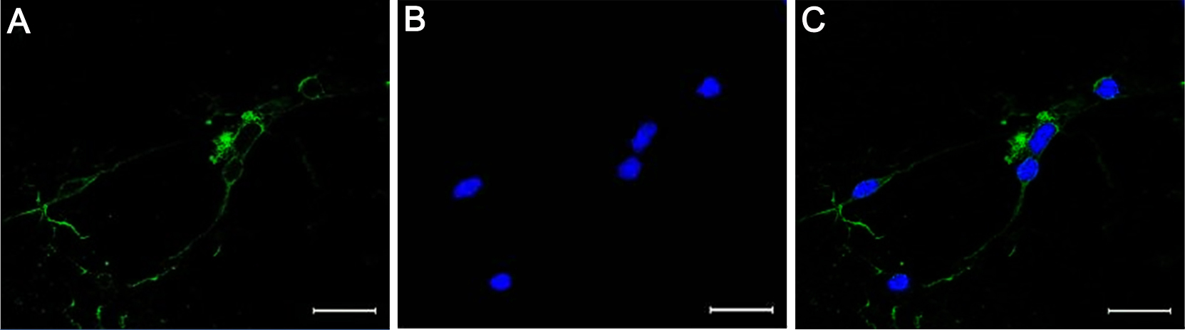
 Figure 1 of
Yang, Mol Vis 2013; 19:1901-1912.
Figure 1 of
Yang, Mol Vis 2013; 19:1901-1912.