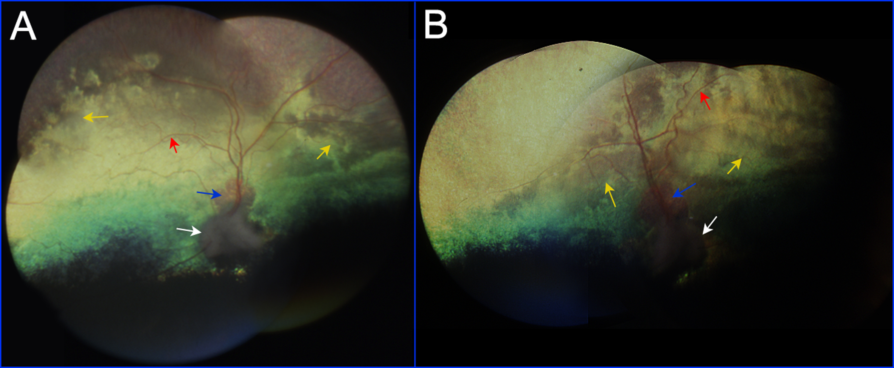
Figure 1. Fundus photographs of a nine-year-old Basenji dog affected with progressive retinal atrophy (PRA; Dog 6,
Figure 2).
A: Fundus photographs of the right eye were assembled into a montage.
B: Fundus photographs of the left eye were assembled into a montage. Retinal degeneration is evidenced by the thin retinal
blood vessels (red arrows), pallid optic nerve head (white arrows), and an irregular pattern of tapetal reflectivity (yellow
arrows). The yellow region immediately superior to the optic nerve head in both eyes (blue arrows) is an expanded area of
conus (see text).
 Figure 1 of
Goldstein, Mol Vis 2013; 19:1871-1884.
Figure 1 of
Goldstein, Mol Vis 2013; 19:1871-1884.