Figure 7. Unaffected brain sections. A: Exemplary hematoxylin and eosin (H&E) stained brain sections of control (CON) and optic nerve homogenate antigen (ONA) animals
2 weeks after immunization (n=5/group). No cellular infiltrates were noted in sections from either group. B: luxol fast blue/periodic acid–Schiff (LFB/PAS) stained sections were obtained at 2 weeks. Normal myelin was observed in
both groups. C: Representative images were taken from horizontal brain sections of CON and ONA animals 10 weeks after immunization and stained
with H&E (n=9/group). No infiltrates were noted at this later time point. D: LFB/PAS-stained brain sections were obtained at 10 weeks. At this time point, no demyelination was observed and no demyelinating
plaques were noted. In summary, brain sections appeared to be without pathological findings at both time points (scale bar:
50 µm).
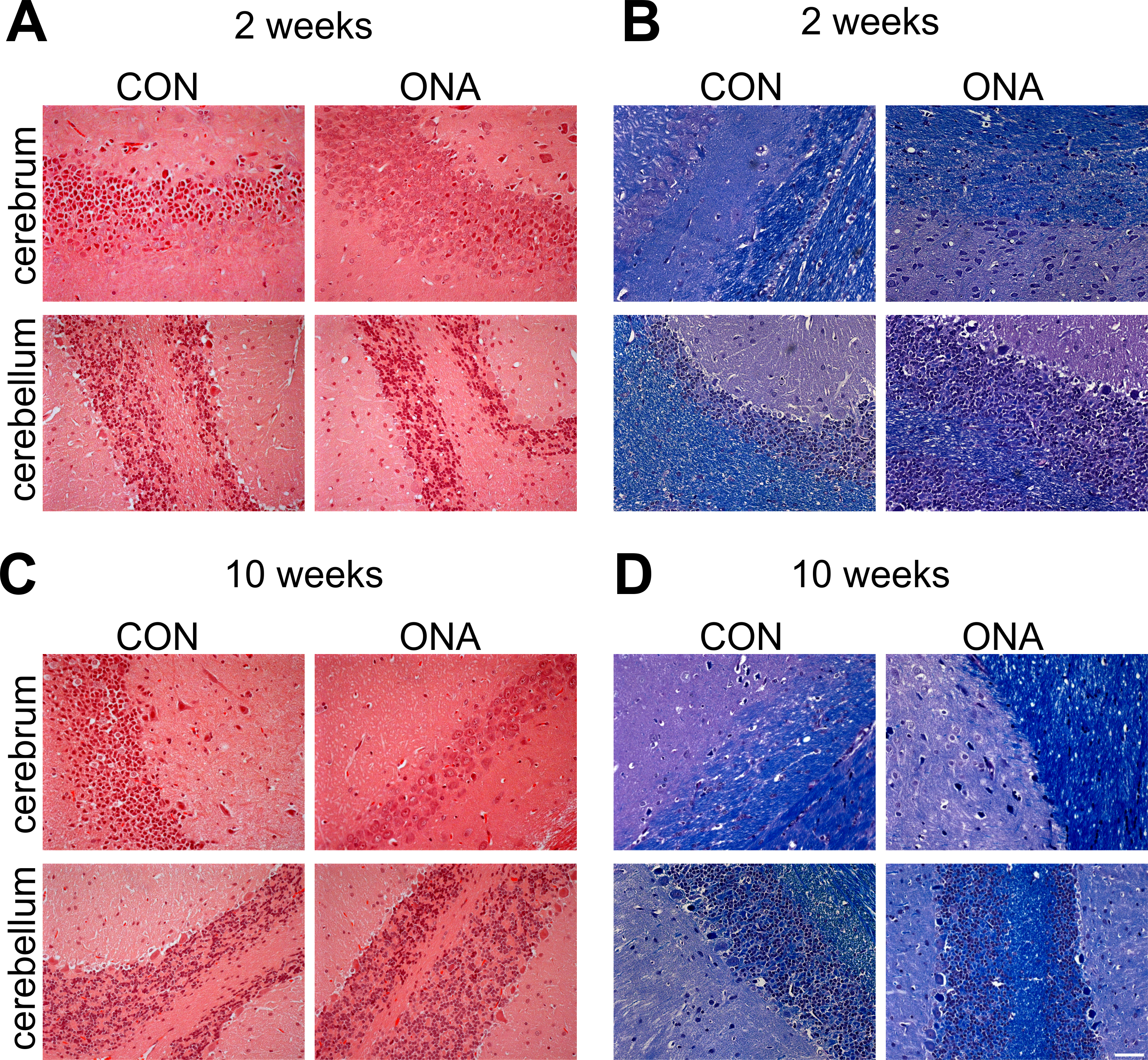
 Figure 7 of
Joachim, Mol Vis 2013; 19:1804-1814.
Figure 7 of
Joachim, Mol Vis 2013; 19:1804-1814.