Figure 6. Optic nerve pathology in optic nerve homogenate antigen animals. A: Longitudinal optic nerve sections from both groups (control [CON] and optic nerve homogenate antigen [ONA]) stained with
hematoxylin and eosin (H&E) showed no inflammatory cells (n=8/group). B: Optic nerve sections were also stained with Nissl. In neither group were cellular abnormalities noted, but the axons of
ONA nerves were more disorganized (n=8/group). C: Luxol fast blue (LFB) staining was applied to assess signs of demyelination (n=8/group). Particularly in the magnified image,
it can be clearly noted that axons of the ONA group are more disorganized, but still appear to be myelinated. D: Nerves were stained with Bielschowsky silver impregnation for axon evaluation (n=8/group). In ONA sections, many more damaged
or swollen axons were noted. In the magnified section, arrows indicate swollen axons and arrowheads injured axons (scale bar
100 µm and higher magnification 10 µm).
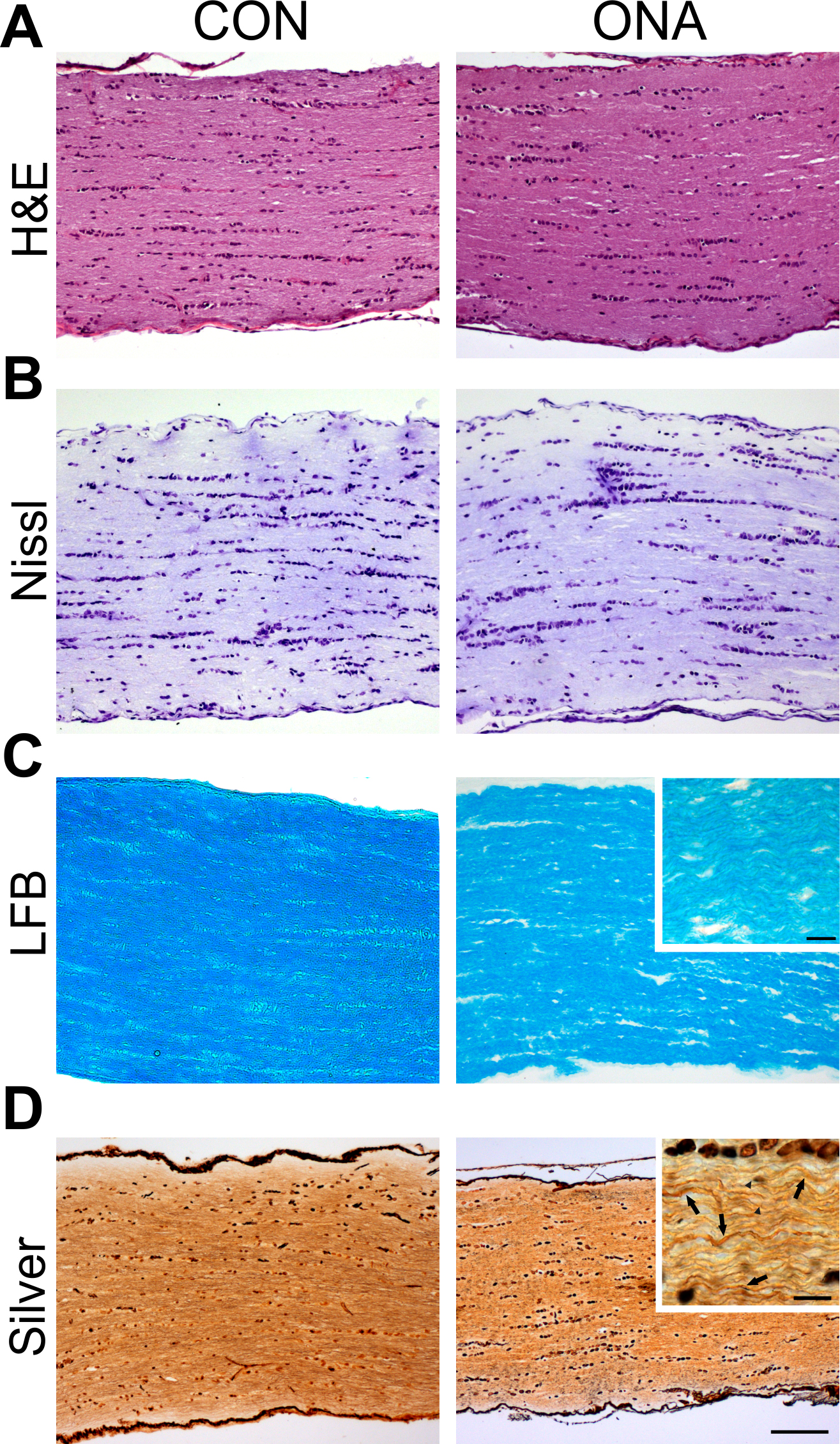
 Figure 6 of
Joachim, Mol Vis 2013; 19:1804-1814.
Figure 6 of
Joachim, Mol Vis 2013; 19:1804-1814.