Figure 4. Development of autoreactive antibodies against the optic nerve. A: Optic nerve sections were incubated with CON or optic nerve homogenate antigen (ONA) serum (n=6/group). No staining was
observed on optic nerve sections before immunization. The staining intensity on sections incubated with ONA serum continuously
increased up to 10 weeks, while few staining signals were observed for CON serum at 4 and 6 weeks and not at the other points
in time. At 4 (p=0.02), 6 (p=0.0003), and 10 weeks (p=0.00001), the ONA group had a significantly higher antibody score compared
to controls. A negative control (right) was incubated only with secondary antibody and did not show any positive staining.
B: Mean autoantibody levels against optic nerve before and up to 10 weeks after immunization. Both groups were compared at
every point in time using the Student t test. C: Higher magnification of nerve section incubated with ONA serum obtained at 10 weeks. Distinct antibody binding was observed.
Values represent mean±standard error of the SEM; *: p<0.05; ***: p<0.001; scale bar in A 100 µm and in C 50 µm).
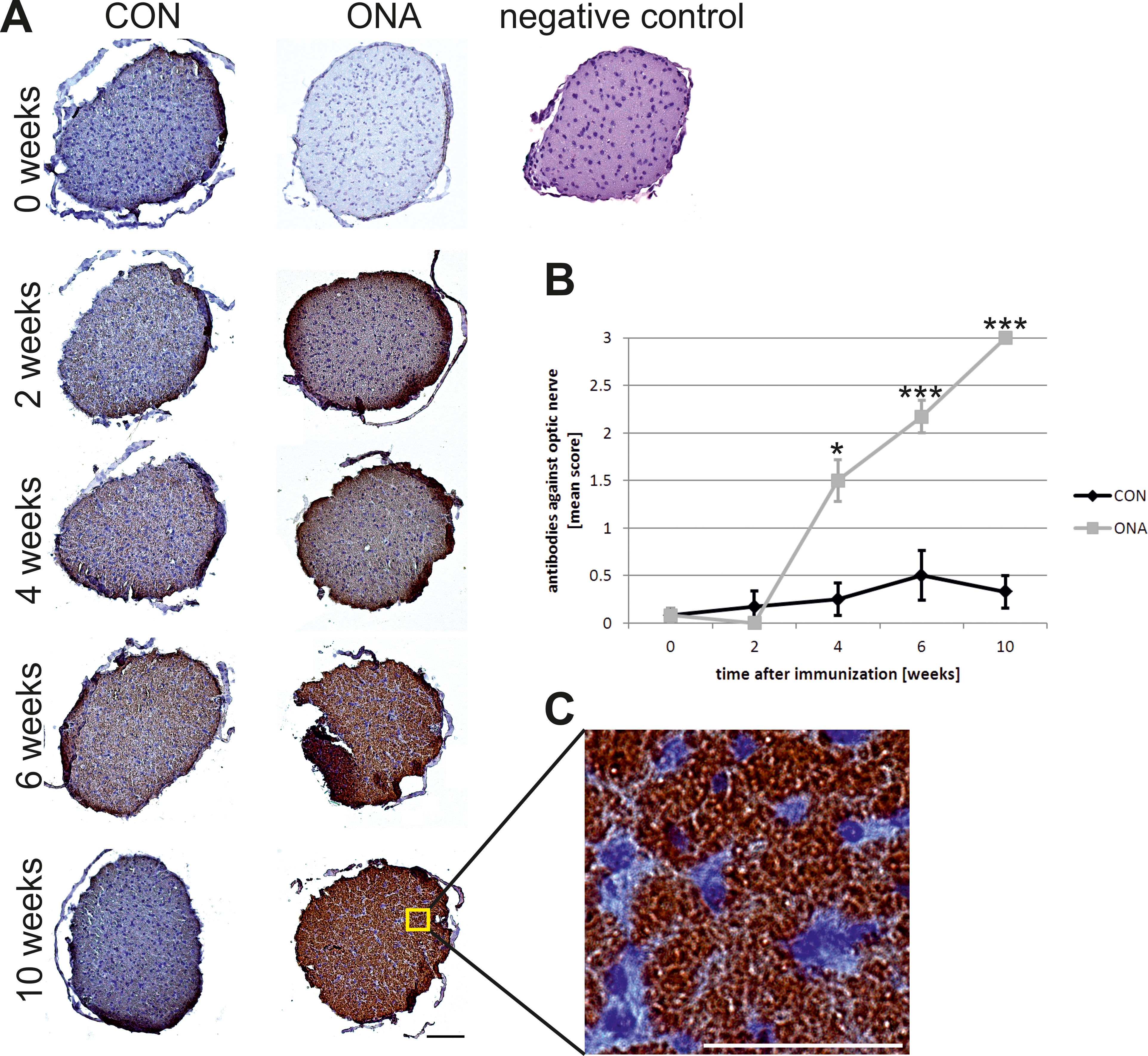
 Figure 4 of
Joachim, Mol Vis 2013; 19:1804-1814.
Figure 4 of
Joachim, Mol Vis 2013; 19:1804-1814.