Figure 2. Evaluation of retinal ganglion cell loss in animals immunized with optic nerve homogenate. A: Exemplary photos of retina cross-sections of the control (CON) and the optic nerve homogenate antigen (ONA) group stained
with the ganglion cell marker NeuN (red) and 4',6-diamidino-2-phenylindole (blue). B: Percentage of loss of NeuN+ and γ-synuclein+ cells in the ONA group compared to controls (n=4 animals/group). Significantly fewer retinal ganglion cells (RGCs) could
be noted in the ONA group compared to CON (NeuN: p=0.00003; γ-synuclein: p=0.0001). The Student t test was performed to calculate the p values. Values represent the mean±standard error of the mean (SEM). Abbreviations:
GCL=ganglion cell layer, IPL=inner plexiform layer, INL=inner nuclear layer (scale bar: 20 µm).
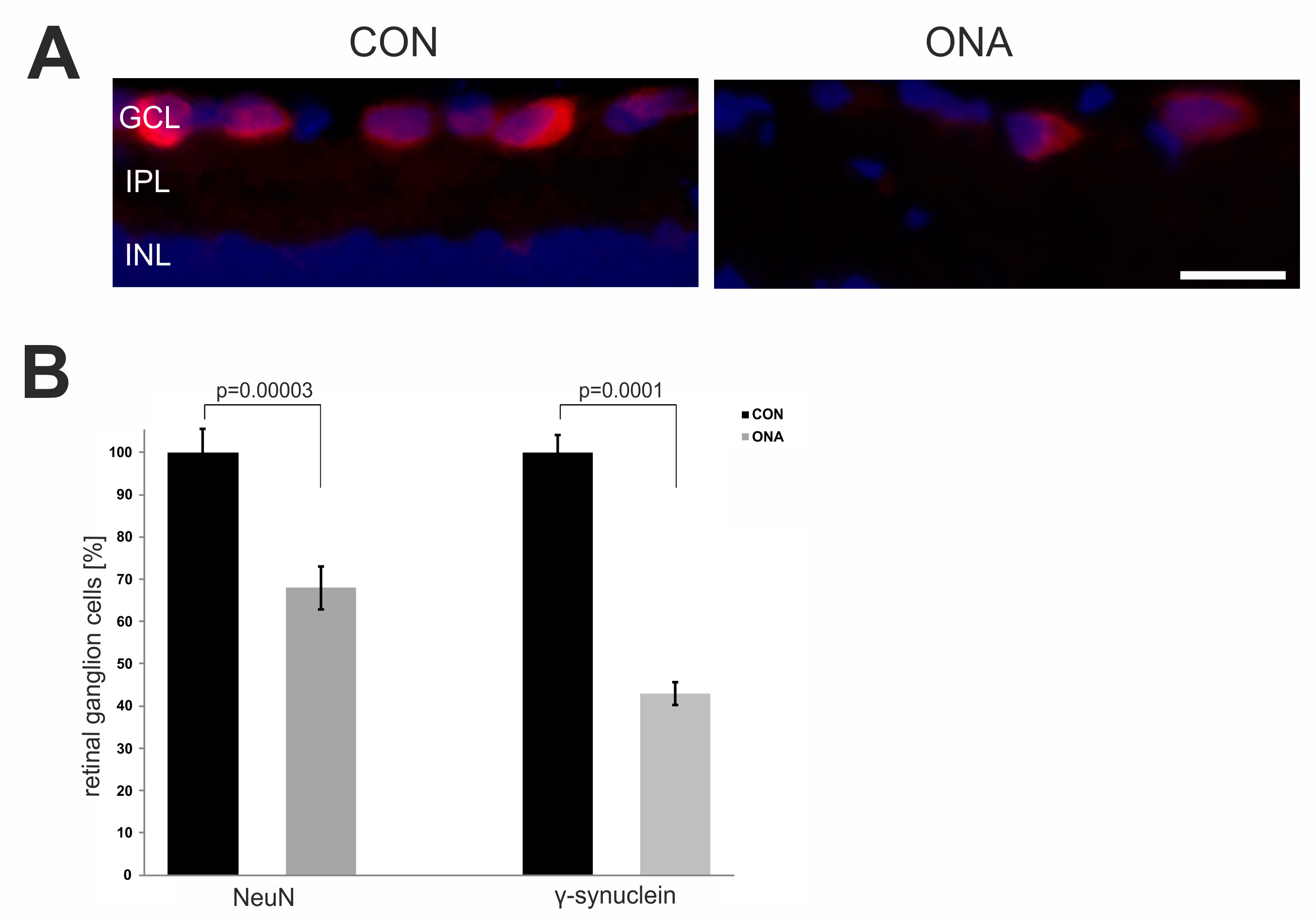
 Figure 2 of
Joachim, Mol Vis 2013; 19:1804-1814.
Figure 2 of
Joachim, Mol Vis 2013; 19:1804-1814.