Figure 1. Intraocular pressure and fundus photos. A: Intraocular pressure (IOP) in controls (CON) and animals immunized with optic nerve homogenate (ONA). IOP was measured before
(=0) as well as 2, 4, 6, and 10 weeks after immunization (n=10 animals/group). No significant difference between groups was
observed (p>0.9 at all points in time). B: Exemplary fundus photos of an ONA animal before and 2, 4, 6, and 9 weeks after immunization. No abnormalities could be detected
during the study. Values represent mean±standard error of the mean (SEM).
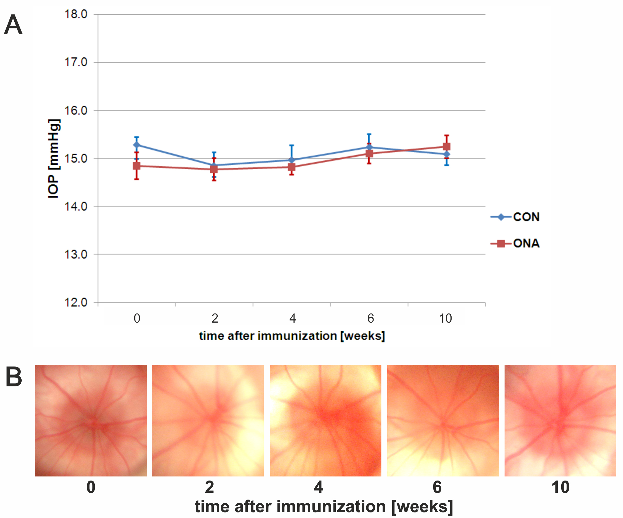
 Figure 1 of
Joachim, Mol Vis 2013; 19:1804-1814.
Figure 1 of
Joachim, Mol Vis 2013; 19:1804-1814.