Figure 4. Expression and non-activation of phospho-p38 and p38 protein in human scleral fibroblasts treated with 1µmol/L all-trans retinoic
acid. Panel A shows expression of phospho-p38 protein in HSFs visualized with indirect immunofluorescence. The nuclei were stained with
propidium iodide dye (red: A1, B1, C1) and the primary antibody was labeled with daylight 488-conjugated secondary antibody (green: A2, B2, C2). Panels A1-A3 shows a negative control incubated in 0.01 M phosphate buffered saline (PBS) with no primary antibody. Panels B1-B3 shows cells incubated in control medium. Phospho-p38 protein is expressed in the cytoplasm and nuclei. Panels C1-C3 shows that there is no change in the expression of phospho-p38 protein in cells incubated with all-trans retinoic acid (ATRA)
for 1 h. The original magnification was 400 X and the scale bar=20 µm. Panel B shows western blot for phospho-p38, p38, and antiglyceraldehyde-3-phosphate dehydrogenase (GAPDH). There is no change of
phospho-p38 or p38 protein after incubation with 1 µmol/l ATRA. The bar graph in panel C shows that there is no effect of ATRA on expression of phospho-p38 protein relative to GAPDH density (n=3). The bar graph
in panel D shows that there is no effect of ATRA on expression of p38 protein relative to GAPDH density (n=3).
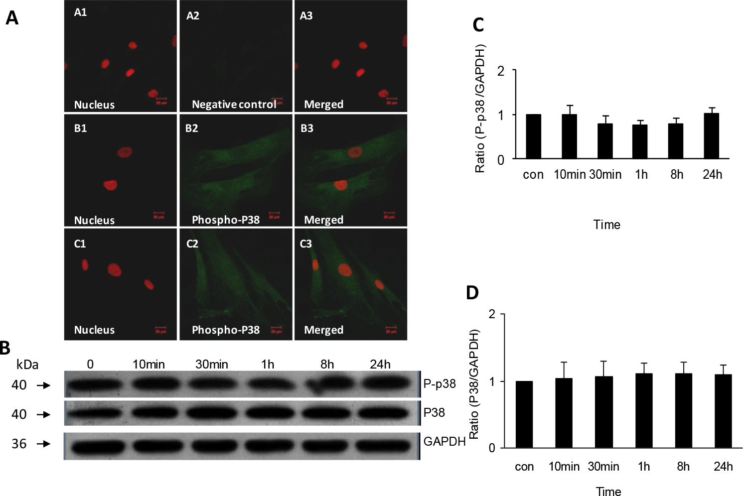
 Figure 4 of
Huo, Mol Vis 2013; 19:1795-1803.
Figure 4 of
Huo, Mol Vis 2013; 19:1795-1803.