Figure 2. Change of expression of phospho-extracellular signal–regulated kinase and extracellular signal-regulated kinase proteins in
human scleral fibroblasts treated with 1 µmol/l all-trans retinoic acid. A shows expression of phospho-extracellular signal-regulated kinase (phosphor-ERK1/2) protein in human scleral fibroblasts
(HSFs) visualized with indirect immunofluorescence. The nuclei were stained with propidium iodide dye (red: A1, B1, C1) and the primary antibody was labeled with daylight 488-conjugated secondary antibody (green: A2, B2, C2). Panels A1-A3 shows a negative control incubated in 0.01 M phosphate buffered saline (PBS) with no primary antibody. Panels B1-B3 shows cells incubated in control medium. Phospho-p44/42 protein is seen expressed in the cytoplasma and nuclei, and the
cells display many protusions. Panels C1-C3 shows cells incubated with all-trans retinoic acid (ATRA) for 1 h. As can be seen, the expression of phospho-ERK 1/2 is
decreased and the morphology of the cells changed. The original magnification was 400 X, and the scale bar=20 µm. Panel B shows western blot for phospho-ERK1/2, ERK1/2, and antiglyceraldehyde-3-phosphate dehydrogenase (GAPDH). There is significant
downregulation of phosphor-ERK 1/2 protein after incubation with 1 µmol/l ATRA from 30 min to 24 h, but no change of ERK 1/2
protein. As can be seen the bar graph of panel C, expression of phospho-ERK 1/2 protein relative to GAPDH density in cells incubated with ATRA is significantly and increasingly
depressed from 30 min to 24 h (n=3, *p<0.05, **p<0.01). In contrast, as seen in the bar graph of panel D, incubation with ATRA did not change expression of ERK 1/2 protein relative to GAPDH density (n=3).
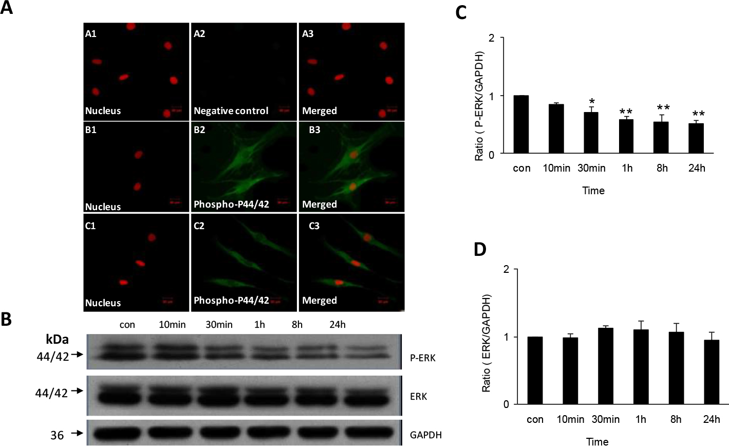
 Figure 2 of
Huo, Mol Vis 2013; 19:1795-1803.
Figure 2 of
Huo, Mol Vis 2013; 19:1795-1803.