Figure 3. Immunofluorescence of adiponectin receptors in the human and mouse radial sections. A: Adiponectin receptor 1 (AdipoR1) was mainly localized at the internal limiting membrane (ILM) and the retinal pigment epithelium
of the retina in humans. B: AdipoR2 expression was not observed in the sections of the human eyeball. C, D: Specific staining was not observed in the sections of the human eyeball in the negative control group. E: AdipoR1 was located in the photoreceptor outer segments and in the internal limiting membrane of the retina in the eNOS−/− mice. F: AdipoR2 was not clearly detected with the fluorescence microscope in the retina of the eNOS−/− mice. G, H: Specific staining was not observed in the sections of the eNOS−/− mice eyeball in the negative control group. Arrows indicate AdipoR1 in the ILM layer. (The calibration bar were 25 μm for
A–B, and 50 μm for C–D. The images were obtained at 400X magnification for A–D and G–H, and 200X magnification for C–F).
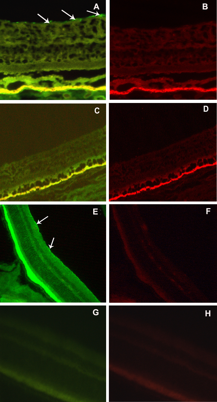
 Figure 3 of
Lin, Mol Vis 2013; 19:1769-1778.
Figure 3 of
Lin, Mol Vis 2013; 19:1769-1778.