Figure 1. Structure of a laser lesion at 14 days post treatment in a mouse model of laser-induced choroidal neovascularization. A: A 1 µm thick plastic section through the center of a lesion was contrasted with toluidine blue. Following initial penetration
through the retinal pigment epithelium (RPE) and Bruch’s membrane (BM), inflammatory signaling occurs and vascular endothelial
growth factor (VEGF) is upregulated to initialize neovascularization. Vessels grow through the break in BM (denoted by the
two white arrows) and ramify between the RPE and the photoreceptor outer segments (red asterisk). B: A close-up of the neovascularized region outlined (red box) in A illustrates that BM (white arrowhead) is compromised (space
between the white arrows). Newly formed vessels have entered the retina through this gap (red arrow) to spread laterally under
the photoreceptors. This mass (red asterisk) displaces the photoreceptors and triggers degeneration (Degeneration Zone). Vessels
within this mass originate from within the choriocapillaris (CC), and not from vasculature within the retina. Note that the
layers of photoreceptor outer segments (OS), inner segments (IS), and outer nuclear layer (ONL) are continuous. C: A confocal image stack of a laser lesion is shown 14 days post treatment. Following removal of the retina, immunohistochemistry
was performed on this flat mount eyecup preparation to highlight endothelial cells (isolectin B4, red), RPE cells (f-actin, purple), and nuclei (blue). The square box shows a top view of the lesion focused at the surface
of the tissue. The horizontal rectangle at the top shows a side view through the image stack at the position of the green
line; the vertical rectangle at the right shows a side view through the image stack at the position of the red line. The blue
line in both the top and side rectangles shows that the image in the large square is from the top of the image stack. Notice
the RPE cells outlined (purple) around the lesion site. Double nuclei are visible in some cells. The thickness of the RPE
cell layer is revealed in the vertical rectangle at the right and a deep capillary is visible within the lesion in the horizontal
rectangle at the top (white arrow head). There is no adhering retinal material above the RPE, indicating that removal of the
retina is complete. The scale bar represent 100 μm. The ganglion cell layer (GCL), the inner nuclear layer (INL), the inner
plexiform layer (IPL), the region of photoreceptor degeneration (Degeneration Zone), and the sclera (Sc) are denoted.
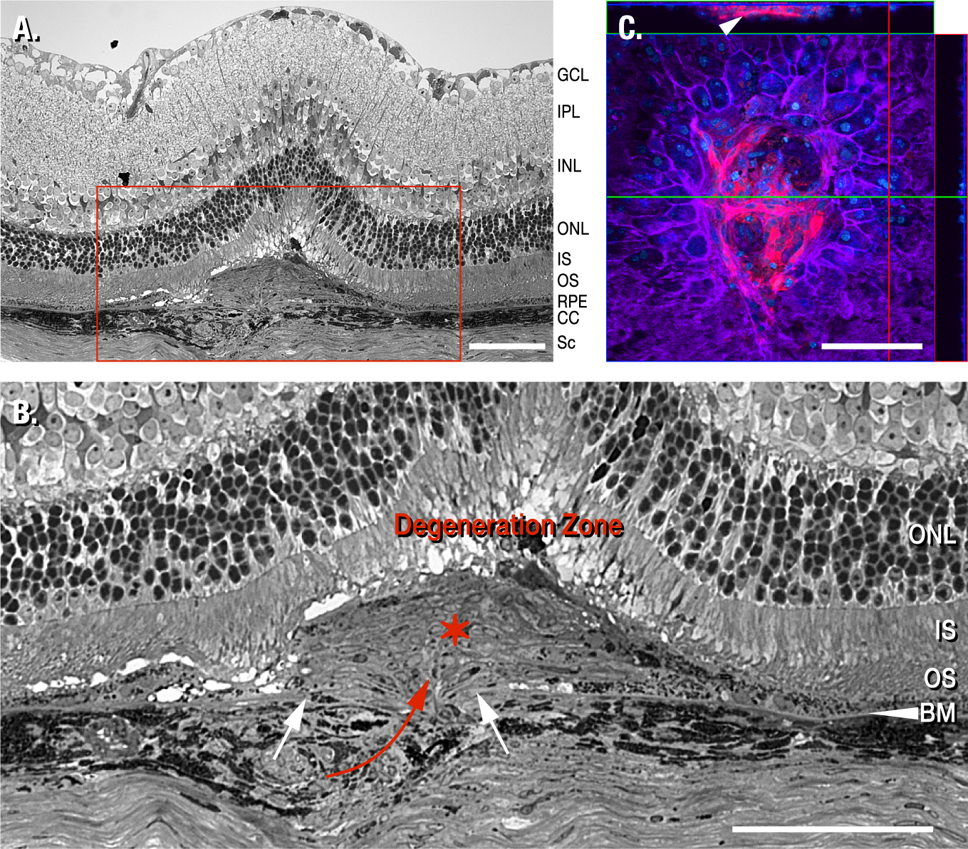
 Figure 1 of
Sheets, Mol Vis 2013; 19:1747-1759.
Figure 1 of
Sheets, Mol Vis 2013; 19:1747-1759.