Figure 3. Pituitary adenylate cyclase-activating peptide (PACAP) binding receptors were expressed in trigeminal nerve (TgN) cells and
lacrimal gland (LG) acinar cells. A: Total mRNA extracted from dissociated TgN cells showed transcripts for the three PACAP receptors. Data are means±SD (n=6).
#Amplification was detected, but it was below the linear limit of detection (LoD). B: Total mRNA extracted from dissociated lacrimal gland (LG) acinar cells also showed transcripts for PACAP receptors. Data
are means±SD (n=4). Positive immunohistochemical staining for PAC1 receptors appeared orange in C: TgN ganglion cells and in D: Schwann cells. This was due to the green PAC1 receptor staining overlaying the red cell-specific staining. E: Higher power magnification showed green dot staining for PAC1 receptors over the red axons. Where blue-staining by 4',6-diamidino-2-phenylindole,
dihydrochloride (DAPI)-nuclei overlaid green areas, this was considered positive staining for the nuclear membrane lamin;
while other green areas without nuclei were considered PAC1 receptor-positive (see Materials and Methods). F: Higher power magnification showed that the green PAC1 receptor staining was eliminated by peptide neutralization of the
primary antibody. G: When primary antibodies for NF and PAC1 were eliminated, only blue-staining (DAPI) nuclei were observed.
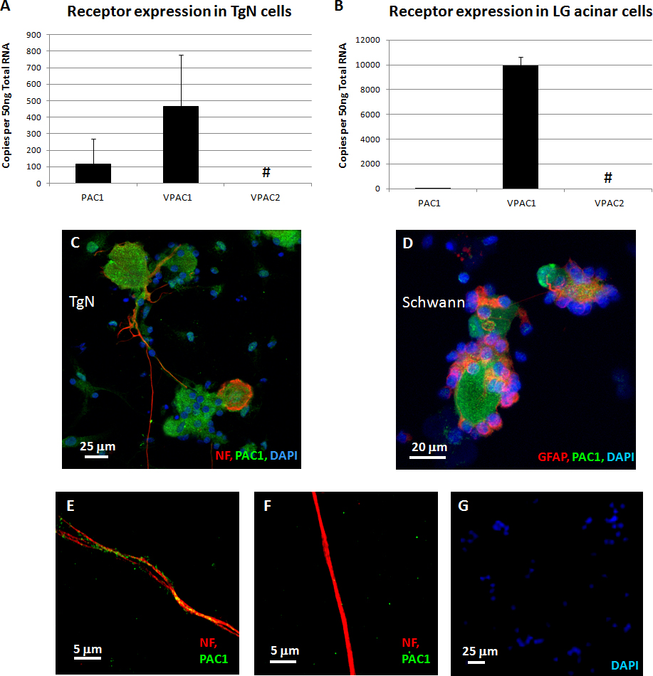
 Figure 3 of
Nakajima, Mol Vis 2013; 19:174-183.
Figure 3 of
Nakajima, Mol Vis 2013; 19:174-183.