Figure 2. Histopathological changes in the retina. A: Hematoxylin and eosin-stained radial sections show morphological alterations with displaced cells (arrows) in 15-month-old
low-density lipoprotein receptor–deficient apolipoprotein B-100-only mice overexpressing insulin-like growth factor II (IGF-II/LDLR–/–ApoB100/100). B: Photoreceptor atrophy (arrows) and thinning of the outer nuclear layer (arrowheads) in outer nuclear layer are also present.
C, D: Normal retinal morphology in 15-month-old C57Bl/6J (C) and a nondiabetic LDLR–/–ApoB100/100 mouse (D). Original magnification in the figure is 400×, and scale bar is 25 μm. Definitions for the abbreviations are: GCL, ganglion
cell layer; IPL, inner plexiform layer; INL, inner nuclear layer; OPL, outer plexiform layer; ONL, outer nuclear layer; IS,
inner segment; OS, outer segment.
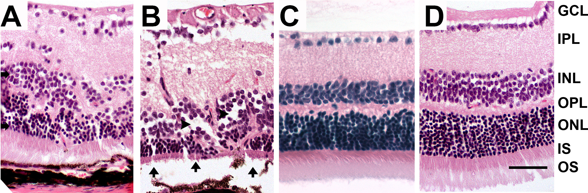
 Figure 2 of
Kinnunen, Mol Vis 2013; 19:1723-1733.
Figure 2 of
Kinnunen, Mol Vis 2013; 19:1723-1733.