Figure 7. Activation of the PI3K/Akt pathway was involved in the protective effect of astaxanthin on ARPE-19 cells. A: Cells were treated with or without 20 µM astaxanthin (AST) for 24 h and then treated with or without 10 µM LY294002 for
30 min before incubation with or without 200 µM hydrogen peroxide (H2O2) for 24 h. Western blot analysis was done using the corresponding antibodies. Cytosolic fractions were immunoblotted with
anti-protein kinase B (anti-Akt), anti-p-Akt, and anti-cleaved caspase-3 antibodies. Nuclear fractions were immunoblotted
with anti-Nrf2 and anti-Lamin B antibodies. B: Quantitative analysis of the relative protein levels in ARPE-19 cells. Data are shown as mean ± standard deviation (SD)
(n=6); *p<0.05 versus control; # p<0.05 versus H2O2 + AST.
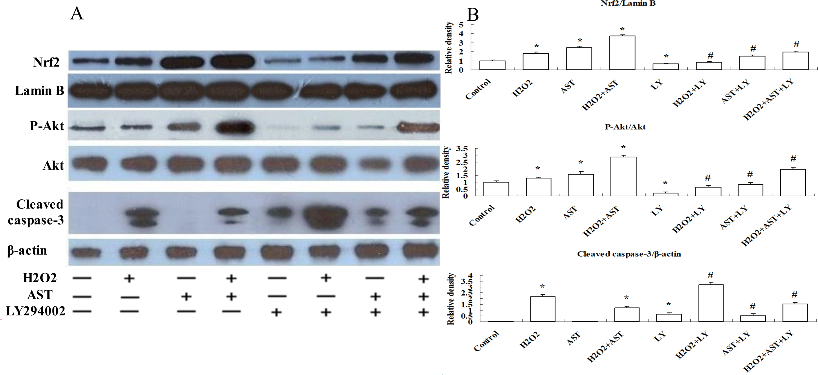
 Figure 7 of
Li, Mol Vis 2013; 19:1656-1666.
Figure 7 of
Li, Mol Vis 2013; 19:1656-1666.