Figure 7. Autophagy assays. A: Western immunoblot with light chain 3 (LC3) antibodies was used to monitor autophagy in human trabecular meshwork (HTM)
cells treated with vehicle control (ctrl), MG132 (5 µM), wortmannin (10 µM), or bafilomycin A1 (1 µM) for 18 h. B–I: Confocal microscopy was used to image HTM cells incubated with control (B, C), MG132 (D, E), wortmannin (F, G), and bafilomycin A1 (H, I). Representative images of immunostaining with the ankyrin repeat and suppressor of cytokine signaling (SOCS) box containing
protein-10 (ASB10) antibody alone (red; B, D, F, H) or colocalized with LC3 (green; C, E, G, I). Nuclei were stained with 4',6-diamidino-2-phenylindole dihydrochloride (DAPI). Scale bars=20 µm. J: The number of ASB10-stained structures (large, small, and total) per nuclei was counted for each treatment. Note that for
bafilomycin A1 the number of small vesicles was an approximation as there were too many to count accurately. Error bars are
the SEM. *, p<0.05 with ANOVA. K: MG132 was applied to human anterior segment perfusion culture. A significant decrease in outflow rate was observed by 3
h after application compared to the vehicle control-treated eyes. Data are the average±SEM. * p<0.05 with ANOVA.
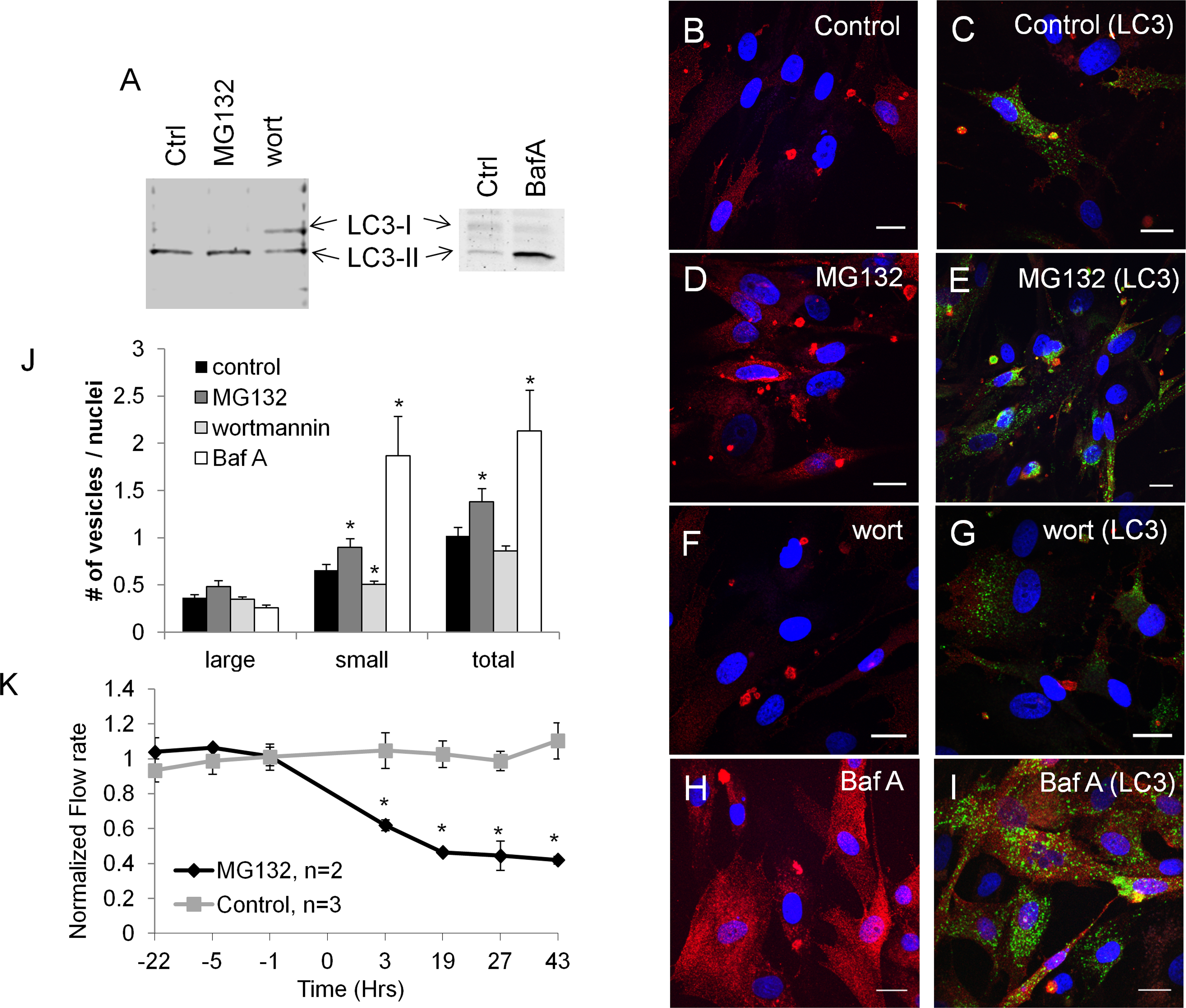
 Figure 7 of
Keller, Mol Vis 2013; 19:1639-1655.
Figure 7 of
Keller, Mol Vis 2013; 19:1639-1655.