Figure 2. Characterization of ankyrin repeat and suppressor of cytokine signaling (SOCS) box containing protein-10-stained structures.
A: The area of ankyrin repeat and suppressor of cytokine signaling (SOCS) box containing protein-10 (ASB10)-stained structures
was measured using FIJI software from 25 confocal images of human trabecular meshwork (HTM) cells derived from four individuals.
One microscope field shows examples of a large structure and small vesicles. Scale bar=20 µm. B: Efficacy of ASB10 knockdown by sh177 lentivirus was determined. Quantitative PCR (qRT–PCR) was used to measure ASB10 mRNA
levels in HTM cells infected with 106 pfu of sh177 or shControl (shCtrl) lentivirus. Values were normalized for 18S RNA. Values are presented as a percentage of
the control±SEM; n=3; * p=0.0001 with ANOVA. Western immunoblot analysis shows significant knockdown of the ASB10 protein
in the HTM cells. C: The sh177 ASB10-silencing lentivirus was applied to human anterior segment perfusion culture at time point 0, and outflow
rate was monitored for a further 93 h. There was a significant (*p<0.001) decrease in outflow rate compared to the control-infected
eyes. Error bars are SEM. D: The number of large (>5 µm diameter) ASB10-stained structures per nuclei in the control HTM and ASB10-silenced HTM cells
was counted. Error bars are the SEM. *, p=0.033 and **, p=0.03 compared to control short, hairpin ribonucleic acid (shRNA)-infected
HTM cells with ANOVA. E: Poisson probability distribution of the large ASB10-stained structures in control and ASB10-silenced HTM cells is shown.
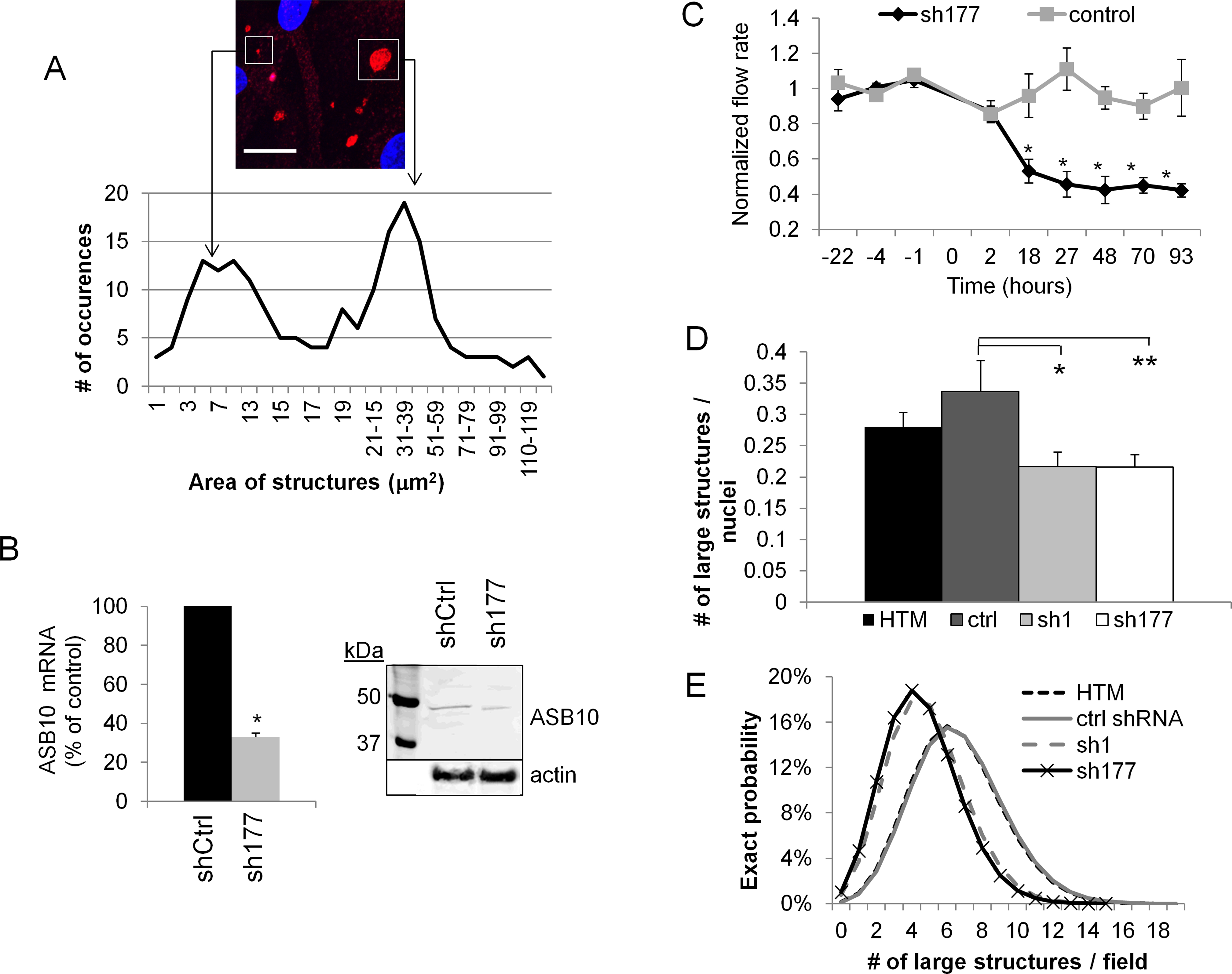
 Figure 2 of
Keller, Mol Vis 2013; 19:1639-1655.
Figure 2 of
Keller, Mol Vis 2013; 19:1639-1655.