Figure 1. Characterization of ankyrin repeat and suppressor of cytokine signaling (SOCS) box containing protein-10 antibodies. A: A schematic diagram of ankyrin repeat and suppressor of cytokine signaling (SOCS) box containing protein-10 (ASB10) shows
the position of the alternatively spliced N-terminus (variant 1 (v1) or variant 3 (v3); green), the ankyrin (ANK) repeats
(olive ovals) and the SOCS box (pink). B: Western immunoblotting was performed to detect endogenous ASB10 in human trabecular meshwork (HTM) cell lysates (left panel)
or 293 cells transfected with recombinant ASB10 variant 3 with a green fluorescent protein (GFP) tag at the C-terminus (right
panel). The control was mock-transfected. Immunoblots were probed with the rabbit polyclonal ASB10 antibody (Rab PAb), the
mouse monoclonal to turbo GFP (tGFP), or the goat polyclonal ASB10 antibody (Gt PAb). Molecular weight markers are shown in
kDa. C–F: Immunofluorescence and confocal microscopy of HTM cells (C, E, F) and normal dermal fibroblasts (D) was performed using the rabbit polyclonal antibody (red, all images) and the goat polyclonal antibody (E, green). A negative control with no primary antibody is shown (F). Nuclei were stained with 4',6-diamidino-2-phenylindole dihydrochloride (DAPI). Scale bars=20 µm.
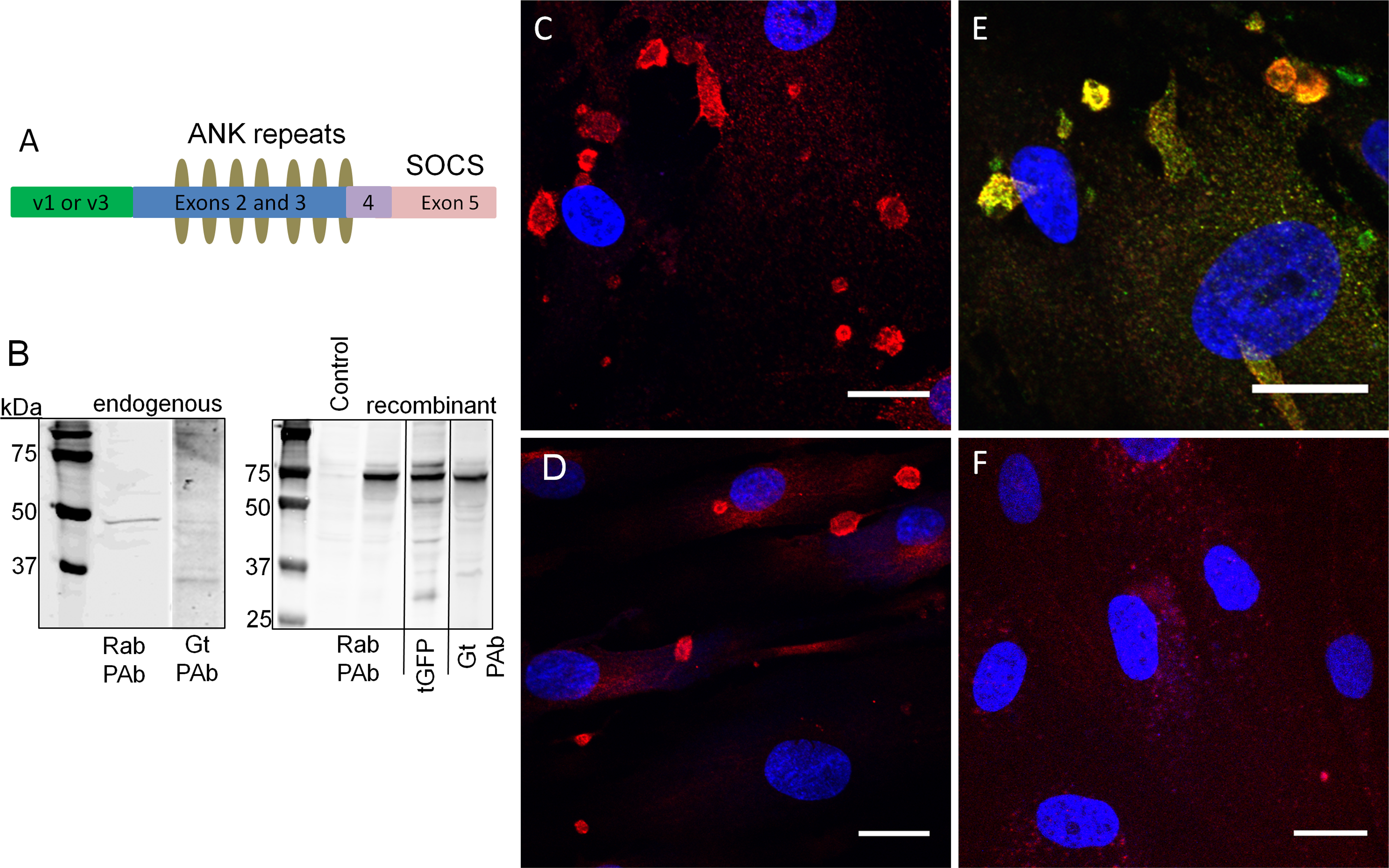
 Figure 1 of
Keller, Mol Vis 2013; 19:1639-1655.
Figure 1 of
Keller, Mol Vis 2013; 19:1639-1655.