Figure 2. Ophthalmologic examinations of the left eye of the proband (III:20) from Family A. Slit-lamp photograph (A) and dilated fundus examination (B) show the floccular turbid of the vitreous body. B-mode ultrasonography shows the high echo in the vitreous body (C). Histochemical examination of vitrectomy specimen stained with Congo red shows amyloid deposits (in red; D).
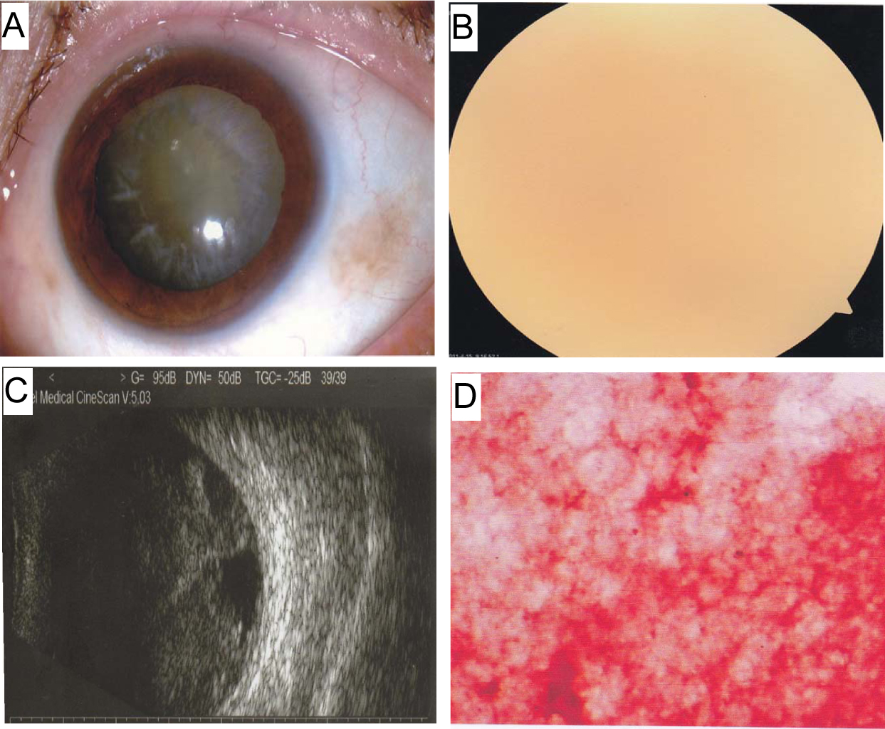
 Figure 2 of
Zhang, Mol Vis 2013; 19:1631-1638.
Figure 2 of
Zhang, Mol Vis 2013; 19:1631-1638.