Figure 5. Analysis of rhodopsin phosphorylated in Ser334 in the outer segment (OS) and pellet (P) of rat retinas exposed to low-intensity light. A: A western blot of rhodopsin (phospho-Ser334) in retinas after exposure to a regular light dark cycle (LD Cycle; 12 h L 200 lx: 12 h D) or to 48 h, 4 or 7 days (LL 48
h, LL4D and LL 7D) of constant light. Experimental animals kept in the dark (DD) are also included. The margin shows the molecular
mass (right). B: Rhodopsin (phospho-Ser334) quantification of OS fraction in retinas after exposure to LD Cycle 48 h (12 h L 200 lx:12 h D), 4, 6, 8, 18, 24 or 48 h
of constant low light (LL 4h, LL 6 h, LL 8 h, LL 18 h, LL 24 h and LL 48 h). Data are means ± SD (n=2 animals/group) from
three independent experiments. One-way ANOVA showed no significant statistical differences (p=0.170). C: Rhodopsin (phospho-Ser334) quantification of OS fraction in retinas after exposure to LD Cycle of seven days (12 h L 200 lx:12 h D) or four or seven
days of constant low light (LL 4D, LL 7D). Data are means ± SD (n=2 animals/group) from three independent experiments, p=0.015
by a one-way ANOVA; *: p<0.05 by Duncan’s post- hoc test (LL 7D versus control in the LD cycle).
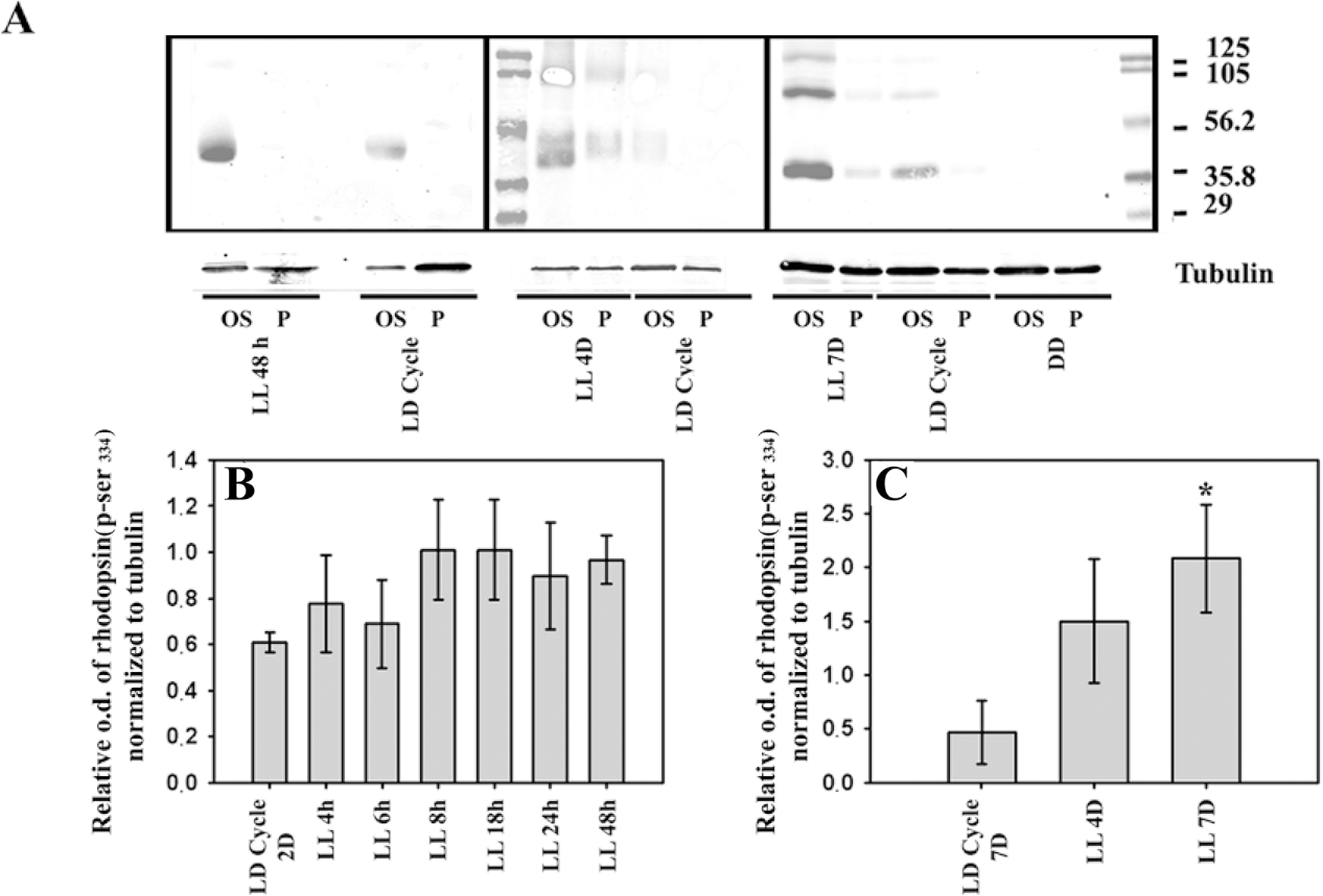
 Figure 5 of
Contín, Mol Vis 2013; 19:1614-1625.
Figure 5 of
Contín, Mol Vis 2013; 19:1614-1625.