Figure 2. Schematic representation of the limbal epithelial crypt. The extracellular matrix composition and structure may regulate limbal
stem cell fate providing information about their position. Depending on the position of cells at the limbal epithelial crypt,
the orientation of the mitotic axis during asymmetric cell division of limbal stem cells could be either vertical or horizontal.
An asymmetric dividing stem cell would give rise to another stem cell and a transient amplifying basal cell that would migrate
to the peripheral cornea when division occurs in the horizontal axis. Conversely, the stem cell could originate another stem
cell and a limbal suprabasal differentiated cell when division takes place following the vertical axis; in this case, loss
of contact between one of the daughter cells and the basement membrane would determine the initiation of the differentiation
process. White arrows indicate the movement of cells after commitment. Differentiation leads to the expression of the terminal
phenotype.
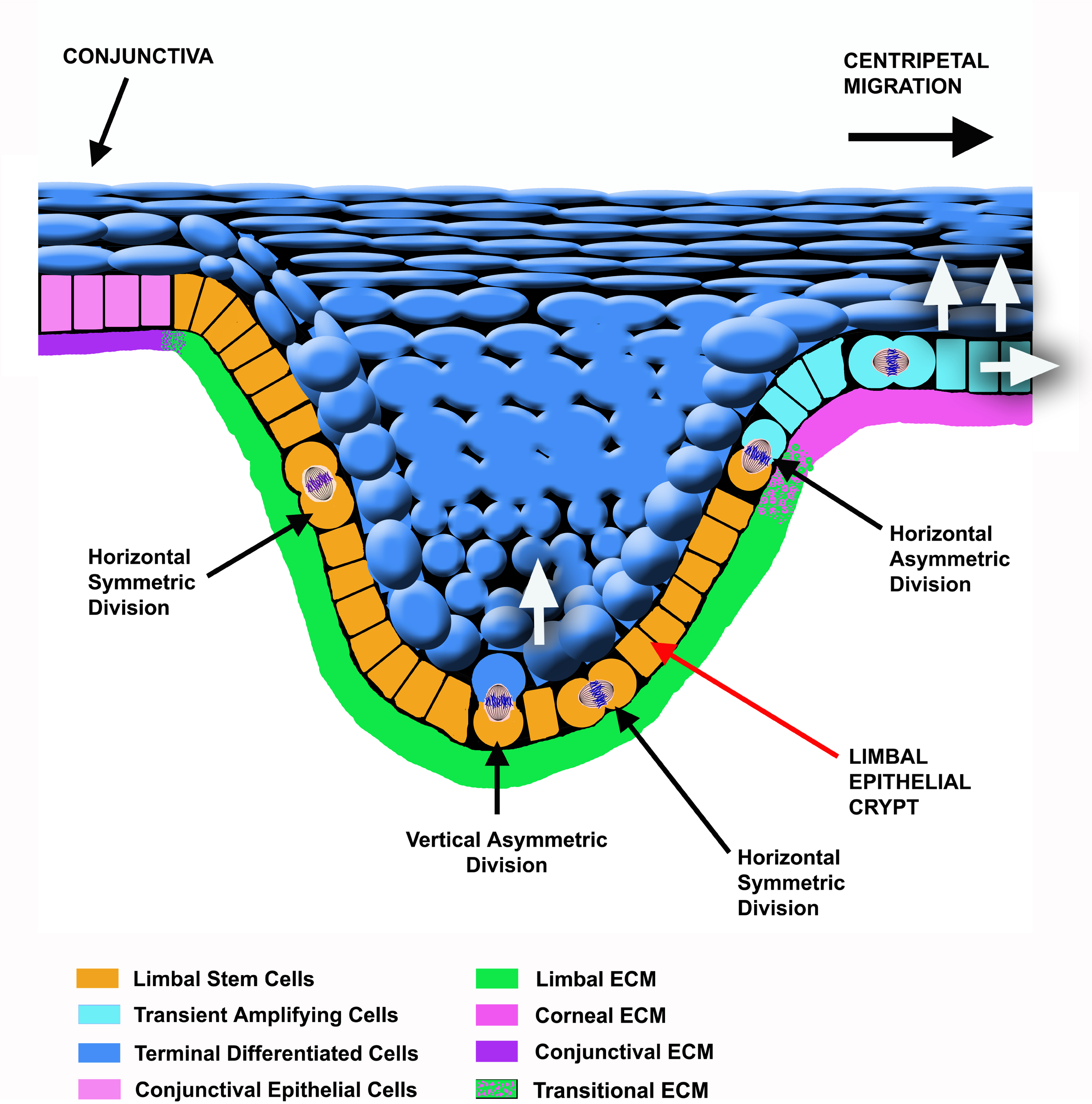
 Figure 2 of
Castro-Muñozledo, Mol Vis 2013; 19:1600-1613.
Figure 2 of
Castro-Muñozledo, Mol Vis 2013; 19:1600-1613.