Figure 2. Isolated bipolar cell with Ca2+ channels in the synaptic terminal labeled with quantum dots (QDs). A: A bright-field image of an isolated bipolar cell. B: A fluorescence image of QDs attached to the synaptic terminal of an isolated bipolar cell. The QD adjacent to the arrow
was located in the focal plane; the dimmer QD above that one was located in a different focal plane. For this experiment,
we used QDs whose emission peaked at 655 nm. The image was averaged from 100 frames acquired at 30 ms/frame. C: To label ribbons, we introduced a fluorescent peptide into the bipolar cell through a whole cell patch pipette. The HiLyte
488-conjugated peptide binds selectively to the CtBP domain of the ribbon protein, RIBEYE. D: Merged image showing that the bright QD overlapped with a region of bright HiLyte 488 fluorescence in the bipolar cell terminal
(arrow).
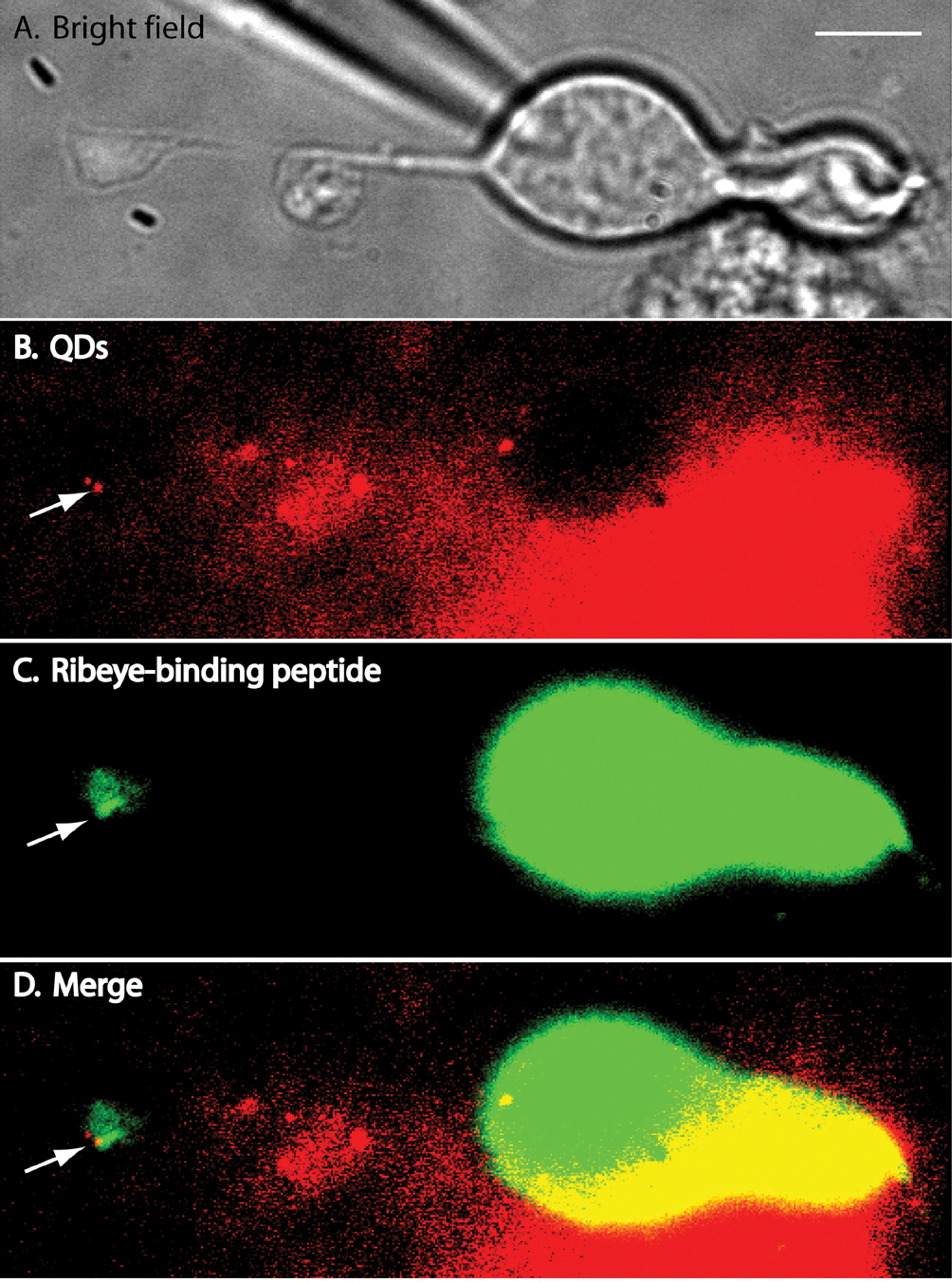
 Figure 2 of
Thoreson, Mol Vis 2013; 19:16-24.
Figure 2 of
Thoreson, Mol Vis 2013; 19:16-24.