Figure 1. Western blot analysis of the anti-α2δ4 antibody. Protein lysates from Ambystoma tigrinum retinas were resolved with western blot analysis. In this blot, we ran increasing quantities of protein lysate in each lane
to optimize the visualization of the presumptive protein bands. Blots were first probed with the anti-α2δ4 antibody, which revealed two distinct bands at 150 kDa and 130 kDa, indicating the presence of the α2δ4 and α2 proteins, respectively. We then stripped the blot and probed it with an anti-β-actin antibody to assess the successive increase
in total protein loaded into each lane.
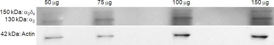
 Figure 1 of
Thoreson, Mol Vis 2013; 19:16-24.
Figure 1 of
Thoreson, Mol Vis 2013; 19:16-24.