Figure 2. Immunohistochemical detection of the presence and location of trypsins. Using the antibody sheep-antitrypsin-1/2/3/4, star-shaped
structures were found in the retina (A) and in the vitreous (B). Similar structures were discovered using the mouse-antitrypsin-2 antibody (C). Double labeling showed colocalization of trypsin-2 (green) and Iba-1 (red), a microglia marker, in the vitreous (D) and retina (not shown). The positive control pancreas was stained after the antibodies sheep-antitrypsin-1/2/3/4 (E) and mouse-antitrypsin-1 (F) were used. The liver showed staining when the mouse-antitrypsin-2 antibody was used (G). The negative controls, lacking the primary antibody, showed no staining (H–J). Panel H is the negative control for sheep-antitrypsin-1/2/3/4, I is the negative control for mouse-antitrypsin-1, and J is the negative control for mouse-antitrypsin-2.
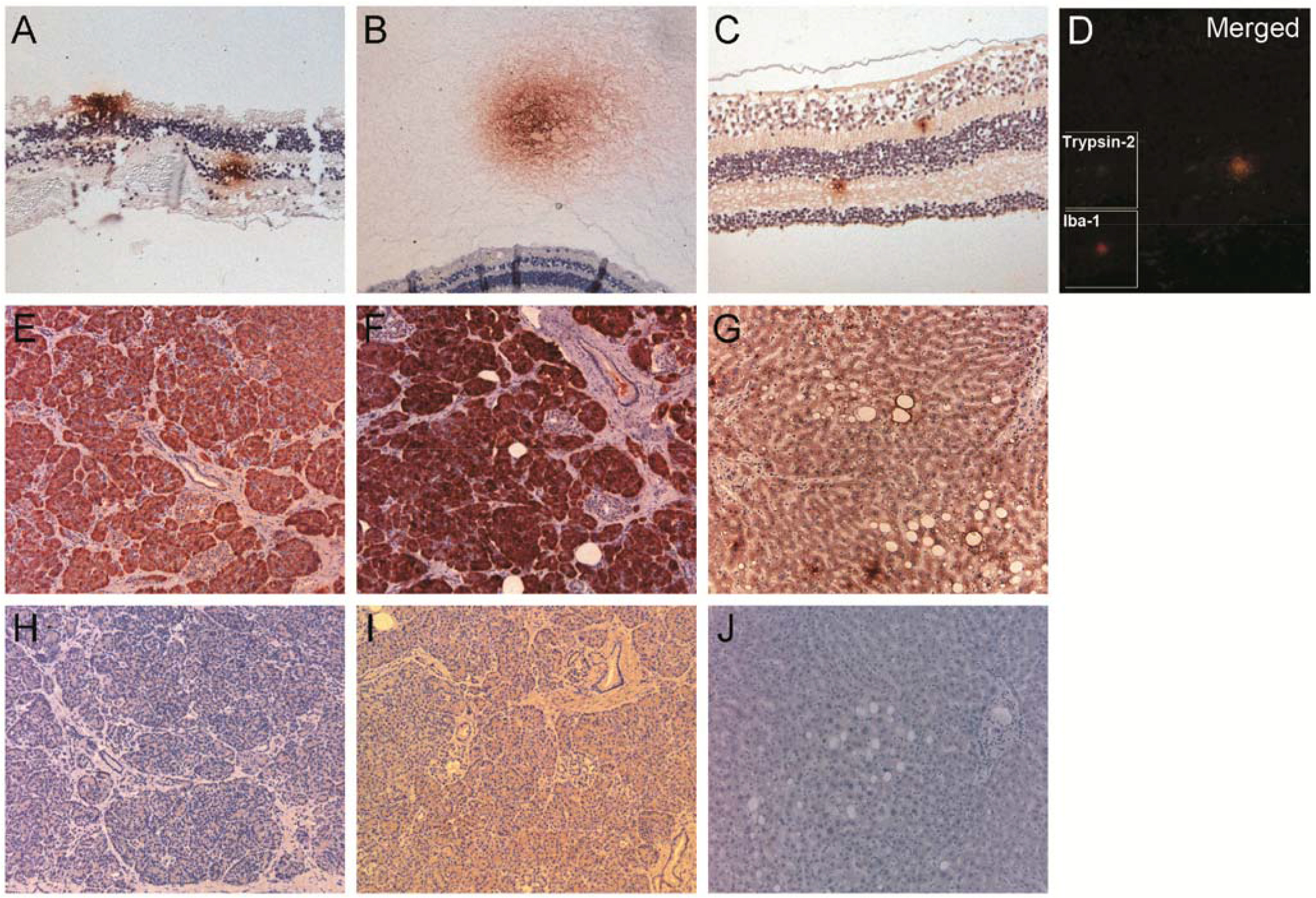
 Figure 2 of
van Deemter, Mol Vis 2013; 19:1591-1599.
Figure 2 of
van Deemter, Mol Vis 2013; 19:1591-1599.