Figure 1. Western blots of vitreous extracts and control tissues incubated with trypsin-1, -2, or -3/4 antibodies. Panel A is concentrated extracts of two vitreous pools (P1 and P2) that were analyzed for the presence of the different trypsin isoforms.
Trypsin-1 was present as a double band and trypsin-2 as a single band (arrowheads). No specific staining was seen after the
antibody against trypsin-3 was used. The left lane contains the molecular weight marker. Panel B shows the positive controls for detecting trypsin, in which recombinant human trypsinogen-1, -2, and -3 were used. Trypsin-1
is present as a double band as well and trypsin-2 and -3 as single bands (arrows). The sizes of all bands were higher than
those detected in the vitreous samples. In all cases, some lighter-stained bands were present. The antibodies used were mouse-antitrypsin-1,
mouse-antitrypsin-2, and rat-antitrypsin-3/4. The right lane contains the molecular weight marker.
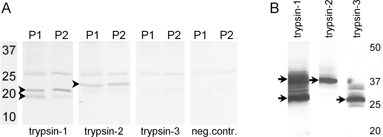
 Figure 1 of
van Deemter, Mol Vis 2013; 19:1591-1599.
Figure 1 of
van Deemter, Mol Vis 2013; 19:1591-1599.