Figure 1. Pedigree and retinal imaging of each patient with potassium channel, subfamily V, member 2 retinopathy. Pedigrees with molecular
status of the three families with potassium channel, subfamily V, member 2 (KCNV2) retinopathy are shown on the left. Retinal images including color fundus photographs, autofluorescence images, and spectral
domain optical coherence tomography are presented on the right. Images of patient 1 (top row), patient 2 (second row from
top), patient 3 (third row from top), and patient 4 (bottom row) are shown.
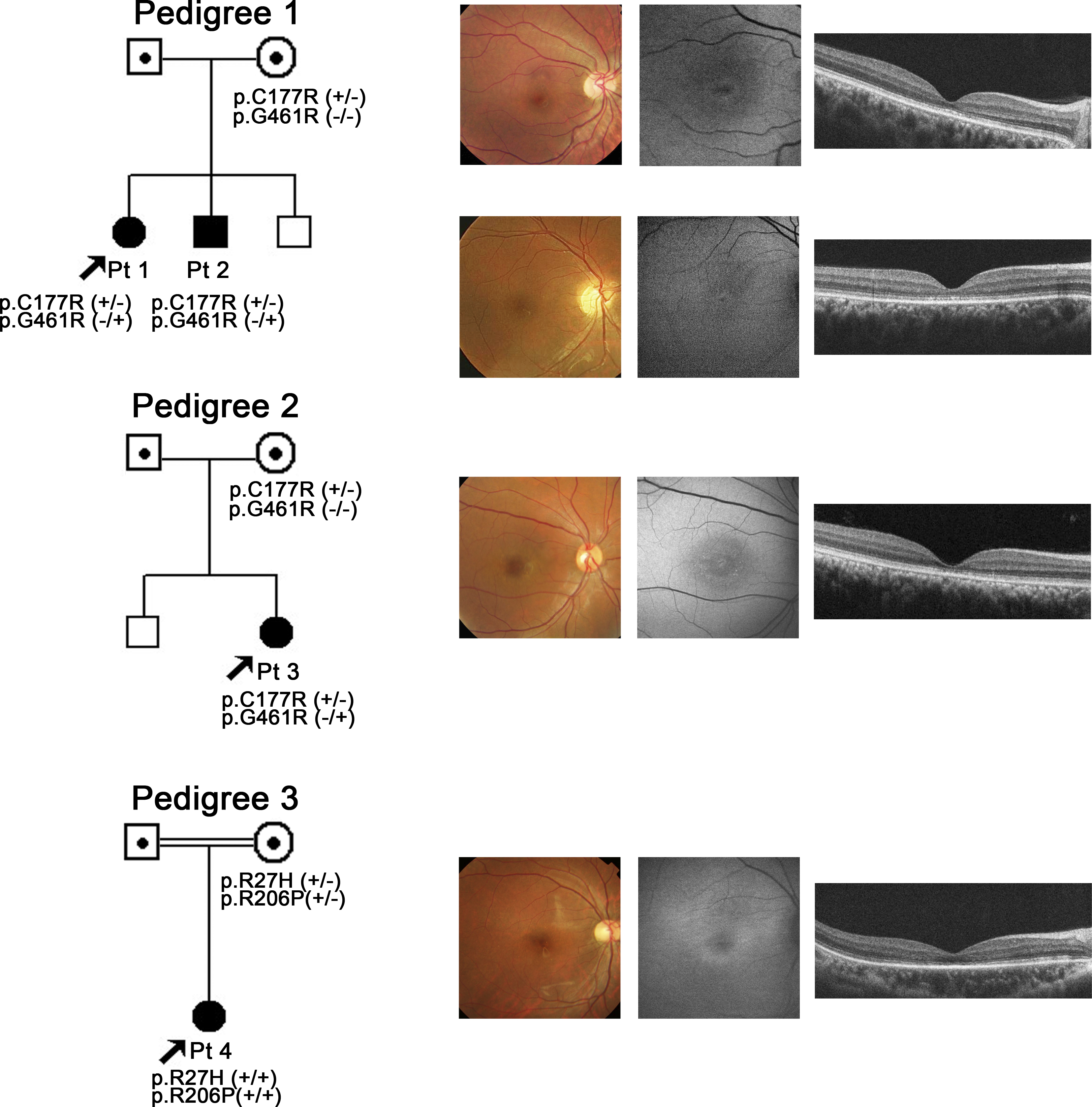
 Figure 1 of
Fujinami, Mol Vis 2013; 19:1580-1590.
Figure 1 of
Fujinami, Mol Vis 2013; 19:1580-1590.