Figure 4. Genomic structure of the EZR exons, domain organization of ezrin and the identified mutation. The red parts indicate the UTR
of the EZR gene, and the blue domains indicate the exons that are translated (upper panel). Ezrin consists of the FERM domain (composed
of three subdomains, designated F1, F2, and F3), α-helical domain, linker region, and C-ERMAD (lower panel). The c.924G>C
in EZR exon 8 and the p.Q308H substitution in ezrin are traced at the bottom of the figure.
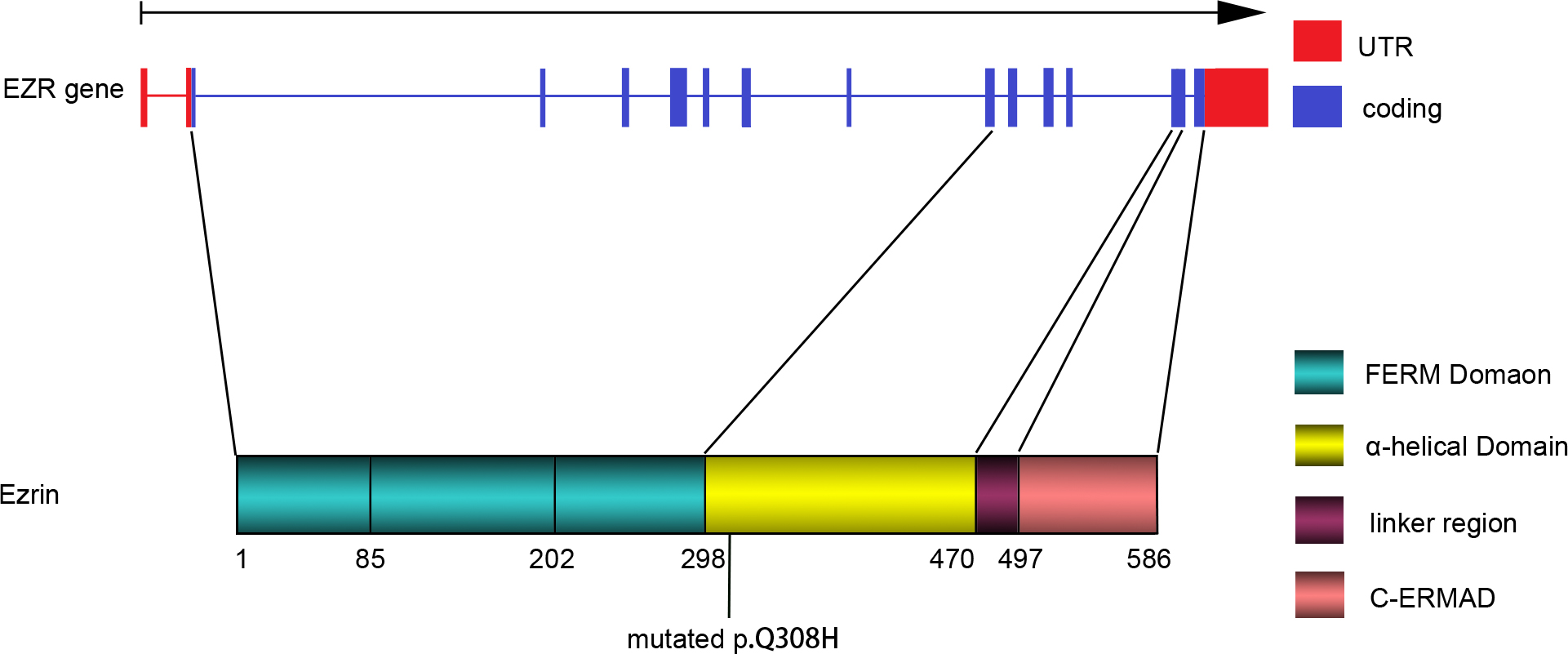
 Figure 4 of
Lin, Mol Vis 2013; 19:1572-1579.
Figure 4 of
Lin, Mol Vis 2013; 19:1572-1579.