Figure 2. Fundus photographs and optical coherence tomography (OCT) of affected individuals from family TB128. A, B: Fundus photographs of individual II:1 at the age of 15 years demonstrate normal optic disc and blood vessels with peripheral
temporal degenerative changes and atrophy of the retina. C: Fundus photograph of individual II:3 at the age of 13 years demonstrates waxy pallor of the tilted optic discs with severe
attenuation of the retinal blood vessels, and bone spicule-like pigmentation in the entire retinal periphery. D: Fundus photograph of individual II:4 at the age of 32 months demonstrates oval optic disc with temporal peripapillary atrophy,
normal color, mild attenuation of retinal blood vessels, and no pigmentary changes in the retina at this stage. E, F: Optical coherence tomography (OCT) of the right and left eyes (RE and LE, respectively) of individual II:1 demonstrates
normal retinal thickness. Macular thickness: RE 274 μm, LE 260 μm. Mean optic nerve fiber layer thickness: RE 101 μm, LE 104
μm. G, H: OCT of RE and LE, respectively, of individual II:3 demonstrates normal retinal thickness. Macular thickness: RE 262 μm,
LE 246 μm. Mean optic nerve fiber layer thickness: RE 105 μm, LE 97 μm.
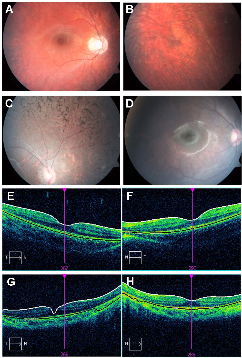
 Figure 2 of
Goldenberg-Cohen, Mol Vis 2013; 19:1565-1571.
Figure 2 of
Goldenberg-Cohen, Mol Vis 2013; 19:1565-1571.