Figure 3. Electron micrograph and digital images of the collagen fibrils of normal, keratoconus (KC), and cross-linked (CXL) corneas.
A: Electron micrograph of collagen fibrils which are present in the normal human corneas. B: Digital image obtained after processing the image shown in A. C: Electron micrograph of the collagen fibrils which are present in the KC corneas. D: Digital image is obtained after processing the image shown in C. E: Electron micrograph of collagen fibrils which are present in the CXL corneas. F: Digital image obtained after processing the image shown in E. The images were displayed by using color coding to demonstrate the distribution of the variable diameters of collagen fibrils.
Collagen fibril color coding: Red=10–15 nm, Green=15–20 nm, Blue=20–25 nm, Yellow=24–30 nm, Aqua=30–35 nm, Pink=35–40 nm,
Brown=40–45 nm.
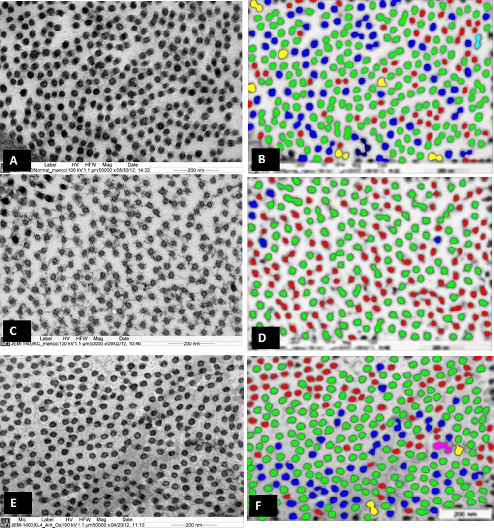
 Figure 3 of
Akhtar, Mol Vis 2013; 19:1526-1537.
Figure 3 of
Akhtar, Mol Vis 2013; 19:1526-1537.