Figure 1. Electron micrograph of cross-linked sample 1 and keratoconus corneas. A: Collagen fibrils are emerging from the posterior part of the Bowman’s layer. B: Interlacing lamellae are present in the anterior stroma. C: Parallel running collagen fibril lamellae are present in the posterior part of the anterior stroma. D: Part of the keratoconus (KC) corneas are showing undulating lamellae. E: Longitudinally running collagen fibrils are decorated with proteoglycan (PGs), in the posterior part of anterior stroma.
F: Cross section of the collagen fibrils are decorated with PGs in the anterior part of the stroma. BW= Bowman’s layer, CF=Collagen
fibrils, L=Lamella, PG=Proteoglycans, S=Stroma.
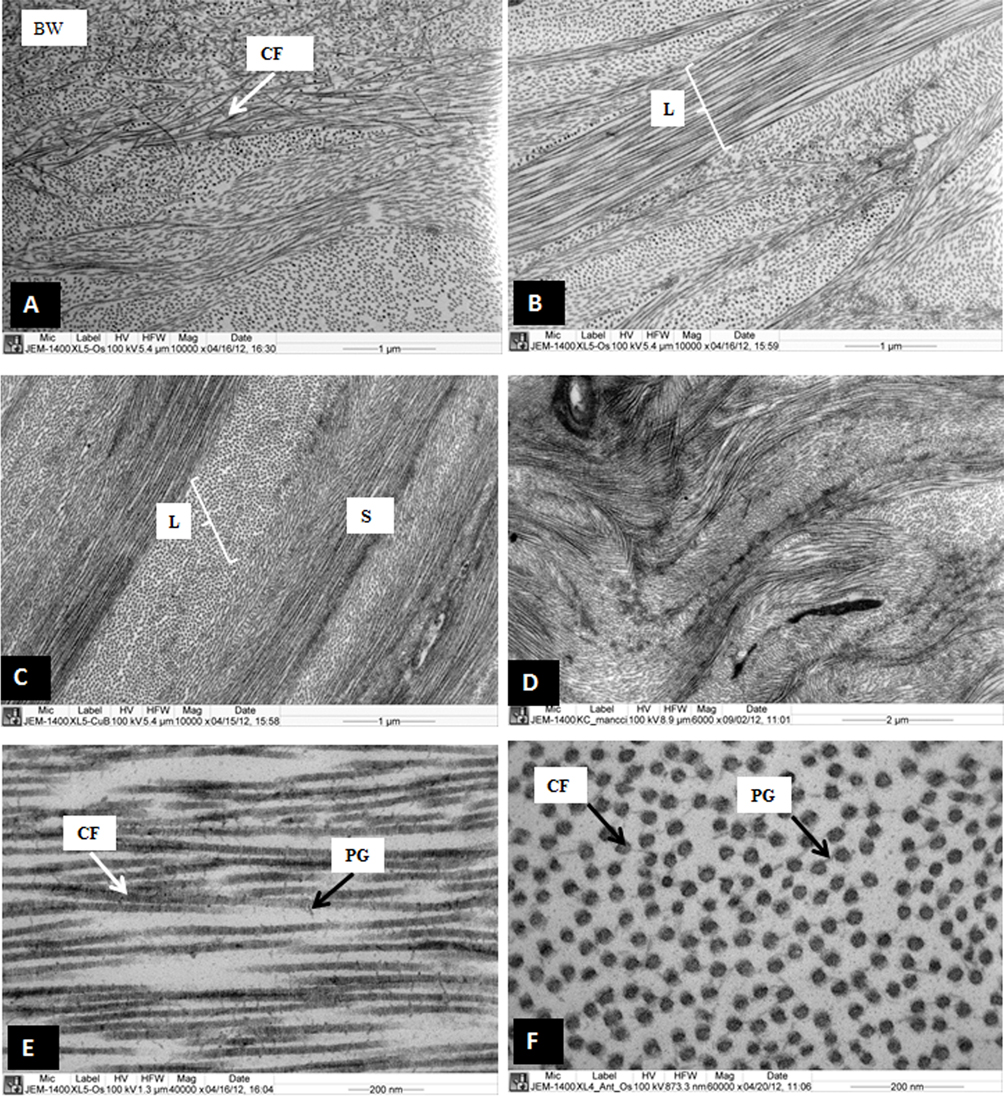
 Figure 1 of
Akhtar, Mol Vis 2013; 19:1526-1537.
Figure 1 of
Akhtar, Mol Vis 2013; 19:1526-1537.