Figure 4. Apoptosis in the infected corneas assessed with flow cytometry. A: The apoptotic ratios in lithium chloride– (LiCl-) versus sodium chloride (NaCl)–treated corneas at 1 and 5 days p.i. were
detected with propidium iodide (PI) staining associated with flow cytometry, as calculated by the average percentage of PI-positive
cells in the infected cornea (D). The respective apoptotic ratios of macrophages (B) and neutrophils (C) in LiCl- versus NaCl-treated corneas at 1 and 5 days p.i. were analyzed with flow cytometry associated with triple staining
for PI, F4/80, and Gr-1, as calculated by the average percentage of PI-positive cells in either F4/80-positive macrophages
(E) or Gr-1-positive neutrophils (F). G: The percentages of the macrophages and neutrophils in the corneal infiltrating cells were determined with F4/80 and Gr-1
staining associated with flow cytometry. Data are the mean±standard error of the mean (SEM) and represent three individual
experiments (n=5). ***, p<0.001.
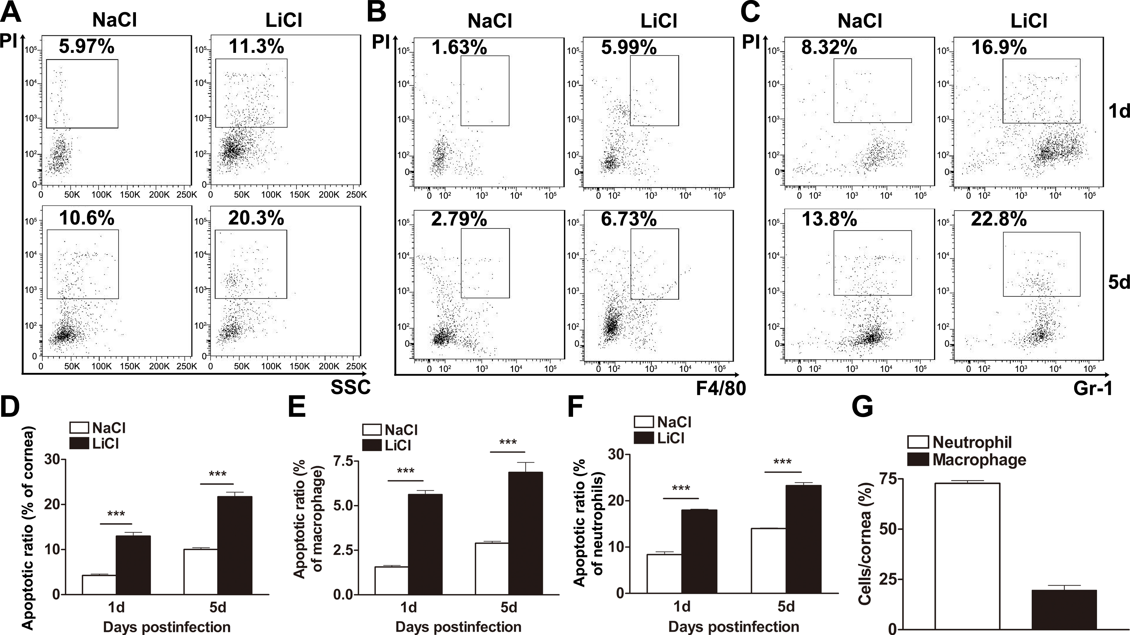
 Figure 4 of
Chen, Mol Vis 2013; 19:1502-1514.
Figure 4 of
Chen, Mol Vis 2013; 19:1502-1514.