Figure 7. Western analysis of retinal proteins following intense white light treatment. Coomassie staining of retinal proteins, with
20 micro grams protein per lane (A), western analysis of oxidative protein markers (B), and the retention of visual cell opsins and arrestins (C) in retinas from rats treated with six hours of intense white light. Light exposed rats, n=4 per treatment, were given zinc
oxide (Z, 1.3 mg/kg), a detergent extract of the Age Related Eye Disease study group (AREDS) vitamin-mineral mix (AR) containing
1.44 mg/kg zinc oxide, rosemary (R, 34 mg/kg), or rosemary plus zinc oxide (R+Z) and compared with vehicle treatment (V) or
no light exposure (D). A: Protein staining was relatively uniform, but increased levels of low molecular weight proteins were seen in lanes 1, 2,
3, and 5. B: Antioxidant treatment (lanes 3–6) reduced the level of carboxyethylpyrrole (CEP) staining for high molecular weight proteins,
while AR and R+Z treatment (lanes 4, 6) also resulted in reduced staining for low molecular weight proteins. Intense light
exposure led to an increase in the level of the Muller cell oxidative stress marker glial fibrillary acidic protein (GFAP)
in all samples (lanes 2–6). C: R+Z treatment was more effective than AR in preserving cone cell opsin and cone arrestin (mCAR) immunoreactivity. Modest
degradation of rod cell opsin and S-antigen, rod arrestin (S-ag) occurred in all light-exposed rat retinal samples. GAPDH
staining, used as an indicator of protein loading, was slightly reduced in lane 3.
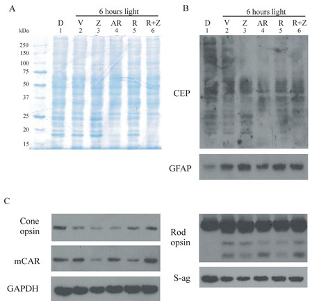
 Figure 7 of
Organisciak, Mol Vis 2013; 19:1433-1445.
Figure 7 of
Organisciak, Mol Vis 2013; 19:1433-1445.