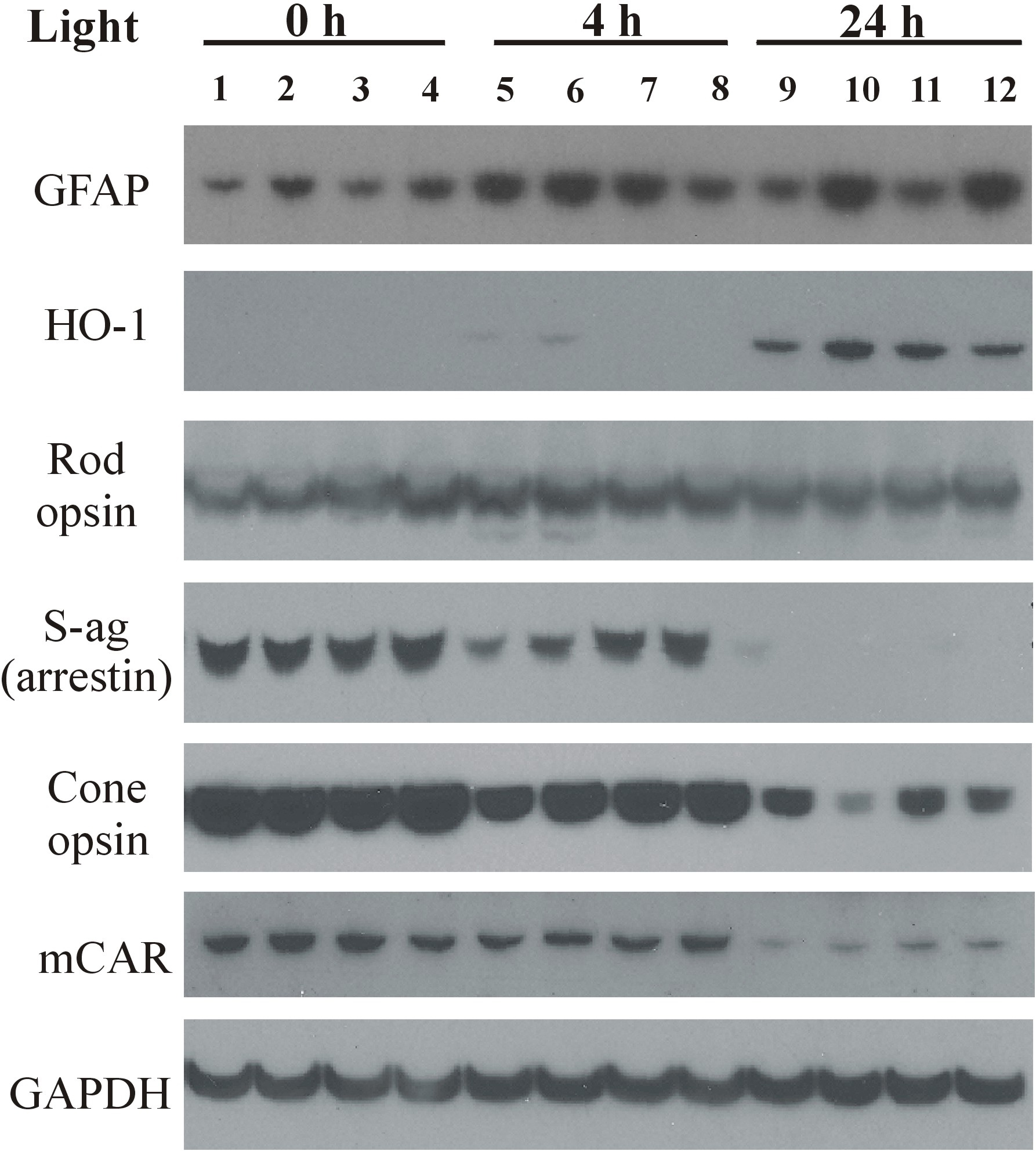
Figure 4. Western analysis following 0, 4, or 24 h of moderate intensity green light. Retinas were excised from dark-reared rats (n=3)
unexposed to light (lanes 1–4), after 4 h of light exposure (lanes 5–8), or from those treated with 24 h of intense light
(lanes 9–12). Retinal proteins from vehicle-treated animals are shown in lanes 1, 5, 9; zinc oxide (0.65 mg/kg)- treated in
lanes 2, 6, 10; rosemary (8.5 mg/kg)- treated in lanes 3, 7, 11 and rosemary plus zinc treatment is shown in lanes 4, 8, 12.
Proteins were extracted for 30 min at 4 °C and then electrophoresed (20 μg/lane) on a 12.5% polyacrylamide gel containing
1% sodium dodecyl sulfate (SDS) followed by transfer to polyvinylidene fluoride (PVDF) membranes and probing with primary
antibodies (see
Table 1). Staining for retinal glial fibrillary acidic protein (GFAP), a marker of Muller cell stress, was increased by intense light
exposure of 4 h duration (lanes 5–8) and by 24 h intense light (lanes 9–12). Zinc treatment also resulted in GFAP induction
(lanes 2, 6, 10), while rosemary appeared to reduce GFAP expression (lanes 3,7,11) compared to vehicle (lanes 1,5,9). Heme-oxygenase-1
(HO-1) levels were elevated by 24 h light exposure (lanes 9–12), while S-ag (rod arrestin), cone arrestin (mCAR), and cone
opsin levels were all lower following 4 or 24 h of light. GAPDH staining indicates that protein loading per lane was relatively
uniform.
 Figure 4 of
Organisciak, Mol Vis 2013; 19:1433-1445.
Figure 4 of
Organisciak, Mol Vis 2013; 19:1433-1445.