Figure 5. Fluorescent photomicrographs showing characteristics of β-III tubulin+ surviving retinal ganglion cells (RGCs) on retinal whole mounts. A: Surviving RGCs in the retinas of normal saline group are shown. The retinas in B, C, and D were all treated with laser-induced IOP elevation. Three weeks after laser-induced IOP elevation (B), the number of RGCs is significantly reduced compared to the control retinas (A). Transfection by control vector AAV-GFP (C) did not affect RGC survival, but overexpression of GAP-43 using AAV-GAP-43-GAP (D) resulted in a substantial decline in RGC viability. Scale bar=50 μm.
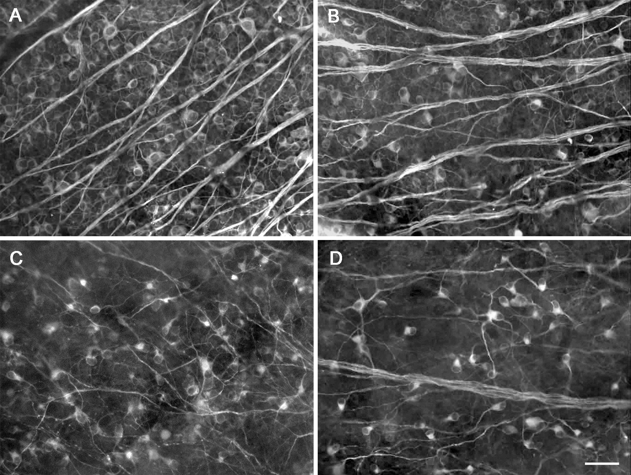
 Figure 5 of
Huang, Mol Vis 2013; 19:1422-1432.
Figure 5 of
Huang, Mol Vis 2013; 19:1422-1432.