Figure 4. Western blots and analysis of growth-associated protein-43 (GAP-43) expression. Data are presented as an adjusted ratio of
GAP-43 to GAPDH. A low level of GAP-43 was expressed in the intact and AAV-GFP groups. With intravitreous injection of AAV-GAP-43,
the expression level of GAP-43 in the optic nerve was increased greatly by more than ninefold compared to the AAV-GFP group
(**p<0.01, n=3 in each group), error bars=standard deviations (SDs).
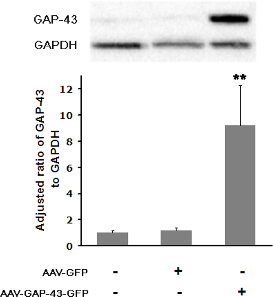
 Figure 4 of
Huang, Mol Vis 2013; 19:1422-1432.
Figure 4 of
Huang, Mol Vis 2013; 19:1422-1432.