Figure 4. Immunofluorescent image of BRN3 positive ganglion cells. A: The merged image of BRN3A (red, panel C), BRN3B (green, panel D) and counterstained with 4',6-diamidino-2-phenylindole (DAPI, blue, panel B) is shown. This double labeling reveals four distinct classes of cell types. A cluster of cells that do not label with either
BRN antibody are indicated with an asterisk. Most cells expressing BRN3 are positive for both proteins (example indicated
with a straight arrow), while a minority express either BRN3A alone (example indicated with a bent arrow) or BRN3B alone (example
indicated with an arrowhead). Size bar=10 µm.
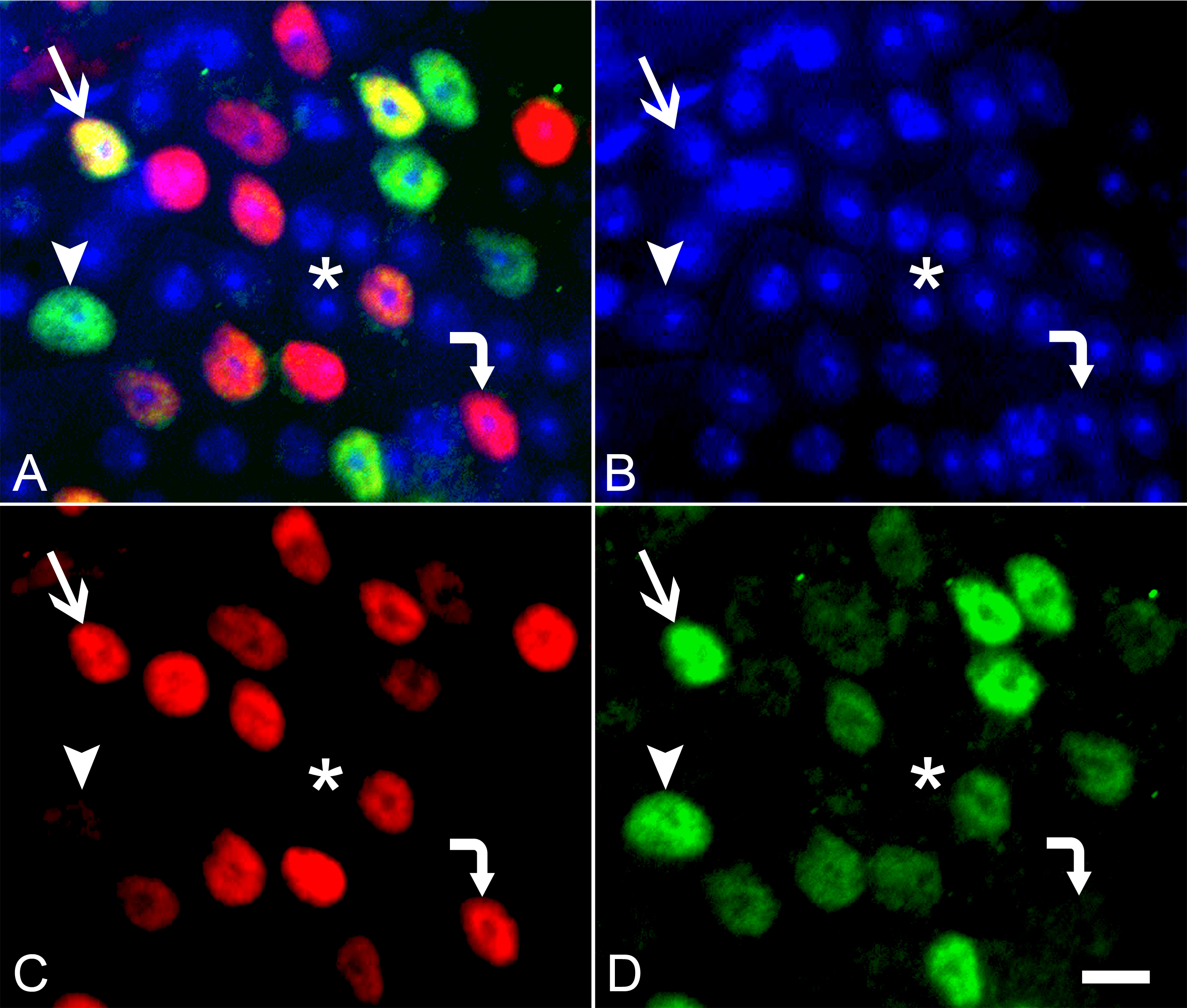
 Figure 4 of
Schlamp, Mol Vis 2013; 19:1387-1396.
Figure 4 of
Schlamp, Mol Vis 2013; 19:1387-1396.