Figure 3. Immunofluorescent image of NeuN labeling of presumptive ganglion cells and cholinergic amacrine cells. A: The merged image of a region of a whole mounted retina from mice expressing tomato fluorescent protein in cholinergic amacrine
cells (red, panel C), immunolabeled for neuronal-specific nuclear protein (NeuN; green, panel D), and counterstained with 4',6-diamidino-2-phenylindole (DAPI, blue, panel B) is shown. Some neuron-like cells do not label with NeuN (exemplar indicated by arrowhead). NeuN colocalizes with the label
for cholinergic amacrines (exemplar indicated with an arrow), in addition to other cell types (asterisk). Size bar=10 µm.
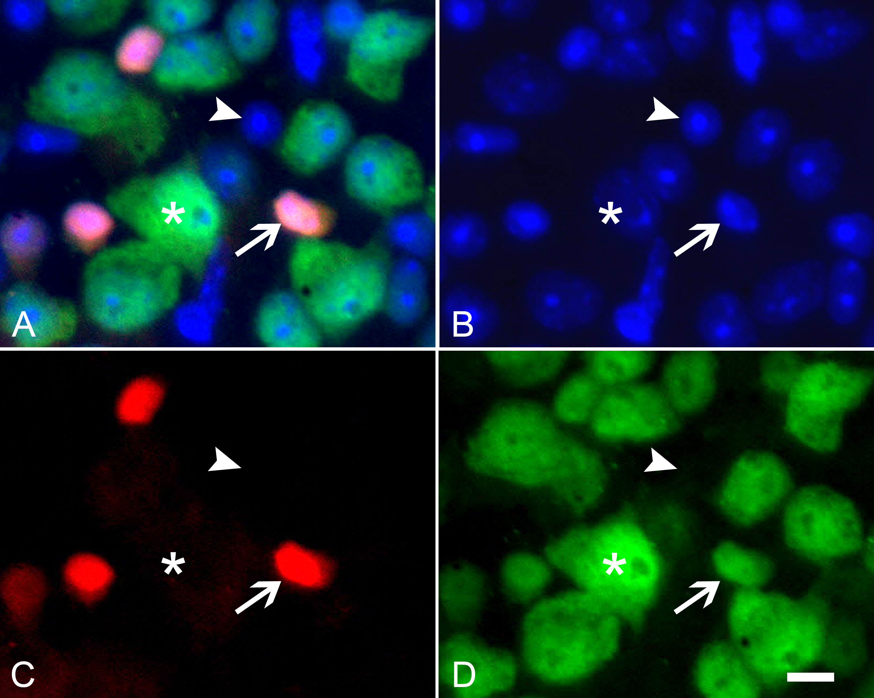
 Figure 3 of
Schlamp, Mol Vis 2013; 19:1387-1396.
Figure 3 of
Schlamp, Mol Vis 2013; 19:1387-1396.