Figure 2. Images of mouse retinas with ganglion cells identified with retrograde labeling. A and B: A plastic section and whole mount of mouse (A) and rat (B) retinas, respectively, are labeled with a mixture of FluoroGold and 4',6-diamidino-2-phenylindole (DAPI, see Methods) stereotactically
injected into the superior colliculus. The section in (A) shows that DAPI (purple) leaks from ganglion cells in the ganglion cell layer (GCL) and penetrates as far as the innermost
layer of the inner nuclear layer (INL), while the outer nuclear layer (ONL) is unstained. FluoroGold (light blue) remains
in the ganglion cells of the GCL. The whole mounted retina shows the distribution of FluoroGold/DAPI positive cells, relative
to the cells stained with DAPI only. These images are electronically enhanced from digitized 35 mm color slide film. C: The whole mount of a retina is stained with retrograde 1,1'-dioctadecyl-3,3,3 3′-tetramethylindocarbocyanine perchlorate
(DiI) label (red, DAPI counter stain). D: The whole mount of a retina is stained with retrograde hydroxystilbamidine (FluoroGold; yellow, TO-PRO-3 counterstain).
In the latter two examples, the retrograde dyes were applied using gel-foam soaked pledgets placed over the exposed superior
colliculus. Size bar in A=20 µm. Size bar in B, C, D=10 µm.
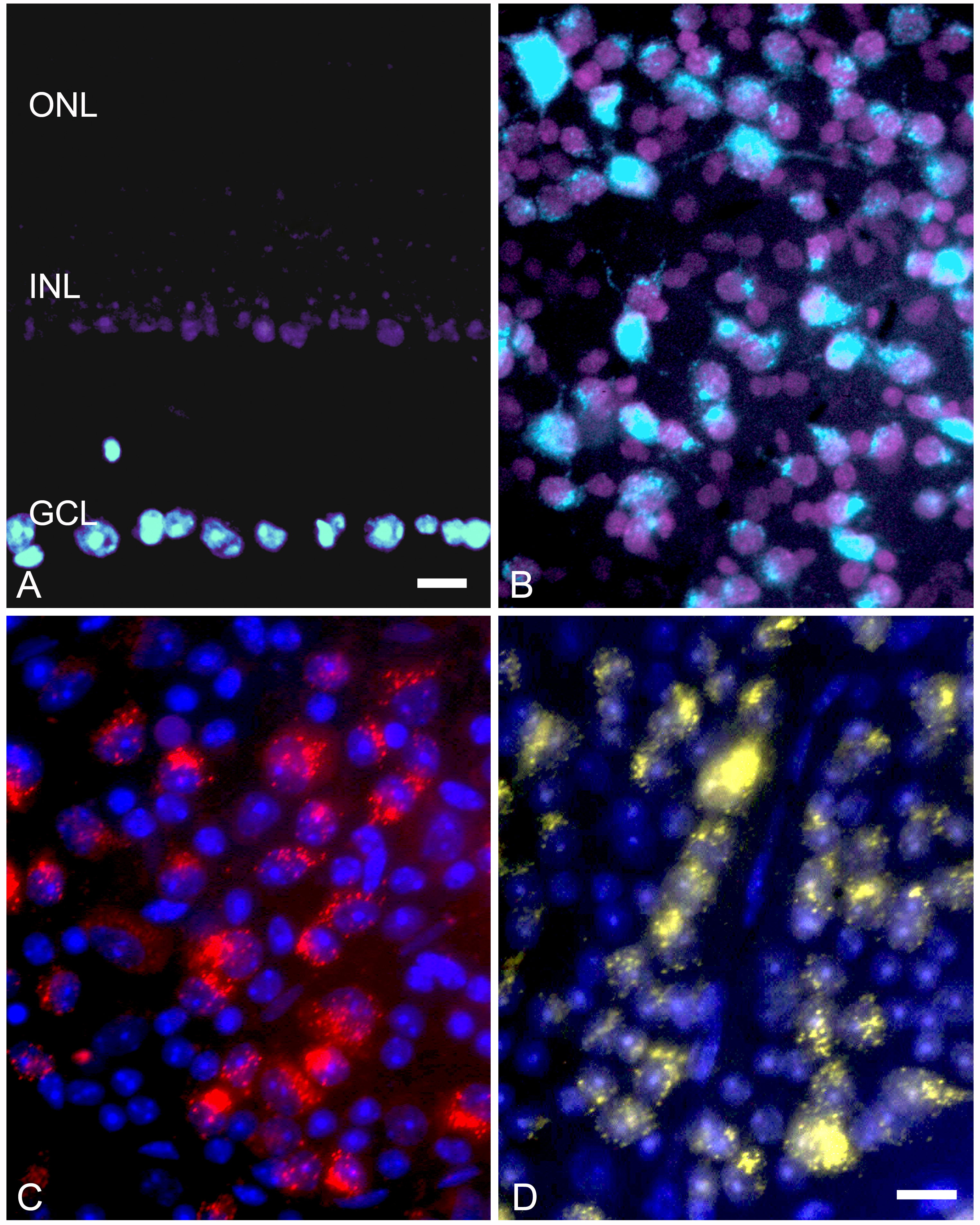
 Figure 2 of
Schlamp, Mol Vis 2013; 19:1387-1396.
Figure 2 of
Schlamp, Mol Vis 2013; 19:1387-1396.